VISITAS RECIENTES

AUTISMO TEA PDF

AUTISMO TEA PDF
TRASTORNO ESPECTRO AUTISMO y URGENCIAS PDF

We Support The Free Share of the Medical Information

Enlaces PDF por Temas

Nota Importante

Aunque pueda contener afirmaciones, datos o apuntes procedentes de instituciones o profesionales sanitarios, la información contenida en el blog EMS Solutions International está editada y elaborada por profesionales de la salud. Recomendamos al lector que cualquier duda relacionada con la salud sea consultada con un profesional del ámbito sanitario. by Dr. Ramon REYES, MD

Niveles de Alerta Antiterrorista en España. Nivel Actual 4 de 5.

Niveles de Alerta Antiterrorista en España. Nivel Actual 4 de 5.
Fuente Ministerio de Interior de España

sábado, 6 de mayo de 2023

Manejo del trauma maxilofacial en emergencia: Una actualización de desafíos y controversias. Management of maxillofacial trauma in emergency: An update of challenges and controversies


Floating in the Face of Danger: MaxFax injuries in the ED

Warning

The content you’re about to read or listen to is at least two years old, which means evidence and guidelines may have changed since it was originally published. This content item won’t be edited but there will be a newer version published if warranted. Check the new publications and curriculum map for updates

Author: Neel Bhanderi / Code: C3AP1d, CMP3, HMP3, EC6, EP2, MaP4, SLO1, SLO3, SLO4, SLO6, TP1 / Published: 19/03/2016

You put an adult trauma call out and start preparing for the patient.

You feel a bit like you have taken the blow yourself, but preparation is key, so get ready.


Preparation

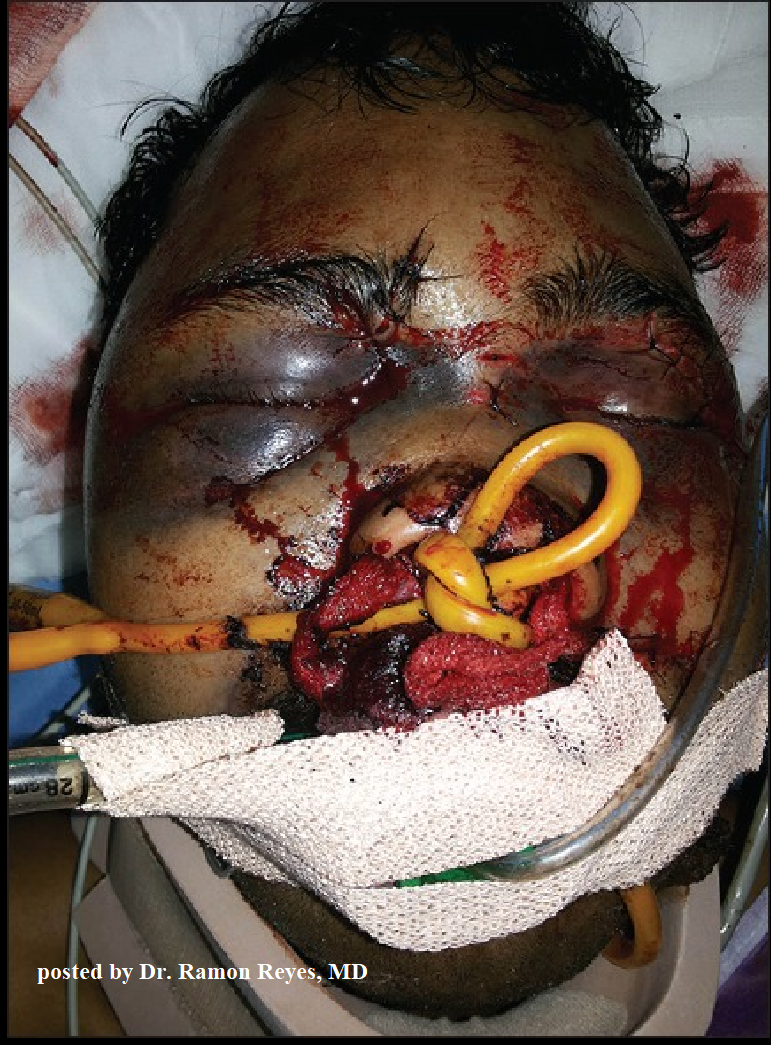
In my experience, facial fractures can bleed and bleed and bleed… Trying to secure the airway can be an anaesthetist’s nightmare. Here’s why:

Its 2 am, they are in an unfamiliar environment (resus) and are often relatively junior!

Significant bleeding (and I mean significant!) from Le Fort and mandibular fractures can obstruct the airway. The blood supply to the face comes from the sphenopalantine and greater palatine arteries (branches of the external carotid artery) as well as the anterior and posterior ethmoidal arteries from the internal carotid artery (that supplies the nasal cavity). That’s a lot of arteries leading to a lot of bleeding!

The loss of normal facial bone structure coupled with the fact that you will have two suction tubes into the mouth in an attempt to clear the blood, will make bag-mask ventilation difficult. (You need two- person technique!)

There will likely be oedema to the soft palate from the mid facial fractures. The patient may also have a traumatic brain injury rendering him unable to self maintain his damaged airway.

My advice is to get your difficult airway trolley into the resus bay.

Action

OK, your trauma team is present and the patient is wheeled in. If he is awake and you are not concerned over a cervical spine injury, sit him upright and forward to allow for postural drainage of the torrential blood flow. Keep him like that until the induction drugs go in, then lie him flat.

What if you are concerned about his C-spine? He comes in supine, with three point immobilisation of his cervical spine. There is a lot of blood. His airway is obstructing.

What can you do?…. Can or should you sit him upright and risk worsening any potential C-spine injury?

If you don’t do something his airway will obstruct.

Take the blocks and tape off and turn him to a left lateral position. This still maintains some immobilisation and also allows for drainage of blood. Get two suction units and remove the rigid yankeur from the end and just use the tubing itself to control the bleeding if it is torrential.

Fracture Assessment

How can you diagnose, by examination only, if the patient has a Le Fort fracture and which one he has?

Stand on the patient’s right hand side. Place your left hand on his forehead to stabilise it. With your right hand, hold the upper teeth and anterior maxilla and gently rock the hard palate.

Le Fort I:

Its a transverse fracture of the maxilla at the level of the nasal fossa. There is separation of the body of the maxilla from the pterygoid plate and nasal septum. Only the teeth and hard palate move.

Le Fort II:

Its a pyramidal fracture through the central maxilla and hard palate. The hard palate and nose move, but not the eyes.

 Le Fort III:

There is craniofacial dysjunction. The entire face is separated from the skull. The fracture line runs through the frontozygomatic suture line, the orbit and base of the nose and ethmoids. The whole face moves.

Le Fort IV: 

Is a Le Fort III along with a fracture to the frontal bone.

Radiopaedia have an excellent blog on the different classifications with great 3D pictures which I really suggest you read.

In Short:

Le Fort I is a floating palate, Le Fort II is a floating maxilla and Le Fort III is a floating face!

In all cases, you can help stop the bleeding by also grabbing the upper incisors and pulling the mid face forward as a temporising measure to disimpact the maxilla.

Back to the case in question: you have now you have diagnosed the patient with a Le Fort III and it is continuing to bleed…..


Airway Management

You need to secure the airway. When I was an ED registrar, chaotic scenes like this, often led to chaotic intubations. I have since learnt a valuable lesson (thanks to a paramedic friend of mine):

“Neel! lets take 10 seconds for 10 minutes…”

Those few words have saved me so many times. Take 10 seconds to gather your team and explain what is about to happen. It will save you 10 minutes of messing around later! Your job is to bring order to chaos! For instance in this situation you could:

Allocate roles (have a very low threshold to  phone the boss on this case).

Set a target oxygen saturation level, that if the patient desaturates to below this you are going to tell whoever is intubating to pull out and ventilate again.

Mark on the neck where the cricothyroid membrane is, just incase you get into a ‘can’t intubate, can’t ventilate’ situation (CICV). Have the surgical airway kit out and open. Read this for revision.

Go through the RSI checklist. Have the DAS failed intubation algorithim to hand.  Listen to HEFT EM Cast for a refresher!

I would have the IGEL or LMA out of the packet ready to insert if needed. Remember Igel size +3 = size of ETT that can be passed through Igel. Realistically if you’re using a size 4 Igel, a size 7 ETT is too tight. Go for a size 6 ETT- bearing in mind you would need a size 10Ch Bougie not the normal 15Ch one!

Get two suction units ready and remove the rigid yankeurs (they tend to get blocked with clots too easily otherwise) and check there is enough battery power in them.

Get your video laryngoscope ready before the RSI (if you are lucky enough have one).

Induce the patient in the left lateral position or sat up right (if no concerns about the cervical spine). Only lay them flat when you are ready to insert the laryngoscope (you can intubate in the left lateral position but it is more difficult.)

Now there are various options on how to intubate this patient. They depend on the time of day, what available staff there are (eg ENT) and what available kit you have.

The options I can think of are:

RSI with direct laryngoscopy

Awake trachesotomy

Awake fiberoptic intubation

Awake direct laryngoscopy with intubation.

LIFTL has a great post on the pros and cons of each.

Lets say the patient is hypotensive and becoming more unconscious, you’ve put out a code red and are pre-loading him with blood. I would use a reduced dose of fentanyl (1mcg/kg) with ketamine (1-2mg/kg- as it gives sedation and preserves the respiratory drive) and rocuronium (1.5mg/kg). I appreciate there may be some arguments about that. I’m not a fan of propofol especially in the unstable trauma patient!


Phew! It all went according to plan and the patient is now intubated! Before you start high-fiving everyone, you need to think about how to control the bleeding.

Controlling the bleeding

It just so happens that your trauma unit has a max fax theatre, which is used once a week for local elective cases. You need to get a pair of epistats and some bite blocks and a semi rigid cervical collar.


An Epistat (bottom left picture) is a nasal catheter which has an anterior and posterior balloon. They are inserted in the same way as a nasopharyngeal airway, along the floor of the nose. The posterior balloon holds 10mls of saline and the anterior balloon holds unto 30 mls. The key to success is inserting an epistat into each nostril first before inflating the balloons. Then simultaneously inject the balloons with saline ( using two people is easier than one person using both of their hands).

The overall aim is to create a rigid vertical structure:

The mandible is stabilised on top of the semi-rigid cervical collar.

The hard palate is held in place against the stable mandible by the bite blocks (top right picture).

The maxilla is fixed in position by the epistats.

There are various accounts on which order this happens. I have always been taught that after the patient has been intubated:

Insert the epistats, like a nasopharyngeal airway, along the floor of the nose. Don’t inflate the balloons yet.

Insert the bite blocks either side of the ETT. There is a groove on them that the ETT should sit next to.

Apply the C-spine collar. (Remember it has been undone for the RSI).

Inject 10 mls of saline to the posterior balloon (white port) of each epistat simultaneously, otherwise you risk disruption of the fractures.

Inject up to 30mls of saline to the anterior port of each epistat (usually green) again simultaneously. I would do this 10mls at a time until haemorrhage control is achieved.

Now you have done all you can to stabilise the patient and transfer on for a CT scan or to the nearest MTC. Your can finally give yourself that deserved high five.


Key learning points are:

Maxfax injuries can have torrential bleeding. Preparation for the intubation is key. Keep the patient upright if they are conscious or in a left lateral position to allow postural drainage.

Team work and good leadership are important. Take time if you need to enable yourself to take control of the case and “reset” everyone before the intubation, so they are all on the same page as you.

Know how to pack the face after the intubation. Remember to inflate the balloons of the epistat simultaneously only at the end.

References:

UK HEMS SOP on max fax bleeding

Radiopaedia: Le Fort Classification

Life in the Fast Lane: Surgical Cricothyroidotomy

Life in the Fast Lane: Airway in Facial Trauma

Difficult Airway Society Guidelines 2015

https://www.rcemlearning.co.uk/foamed/floating-in-the-face-of-danger-maxfax-injuries-in-the-ed/


In any setting ( Austere, Pre hospital, Humanitarian, ED ) , facial fractures can bleed and bleed and bleed… Trying to secure the airway can be a nightmare. Here’s why…

(That’s why we have included a hands on module for this in our workshops , recently adding Rapid Rhino thanks to Louise Mortimer.)


viernes, 5 de mayo de 2023

Narcolepsy / Narcolepsia by JAMA


Narcolepsy causes sleepiness not due to sleep deprivation, poor sleep quality, or sleep apnea. This JAMA Patient Page describes narcolepsy types, signs and symptoms, diagnosis, and treatment. https://ja.ma/3HMJFKL

Hipertensión pulmonar. Infografía by MSP


Hipertensión pulmonar. Infografía by MSP 


#MSPNeumología | Día mundial de la hipertensión pulomar


La hipertensión pulmonar no tratada puede causar complicaciones graves, entre ellas, insuficiencia cardíaca derecha, arritmias cardíacas y muerte súbita.

Conoce la clasificación de la gravedad con esta condición con esta infografía que hemos preparado para ti.

Aprende más en www.revistamsp.com

#MSP: El lugar donde médicos, investigadores y pacientes pueden entrar. #MSPLíderesPioneros

#MSPLegadoQueInspira

SHOCK: Causas, Clasificación y Características Clínicas del Shock. Infografía by MSP


SHOCK: Causas, Clasificación y Características Clínicas del Shock. Infografía by MSP

También leer

8 trampas "ERRORES" a evitar en el control de hemorragia by Dr. Peter Pons, MD FACEP https://emssolutionsint.blogspot.com/2018/09/8-trampas-errores-evitar-en-el-control.html


#MSPGeneral | ¿Sabías qué el shock se produce cuando el cuerpo no recibe suficiente flujo sanguíneo y oxígeno para mantener los órganos vitales funcionando adecuadamente?

Conoce algunas características clínicas de esta condición con esta infografía que hemos preparado para ti.

Aprende más en www.revistamsp.com

#MSP: El lugar donde médicos, investigadores y pacientes pueden entrar. #MSPLíderesPioneros

#MSPLegadoQueInspira


GUIA DE OXIGENOTERAPIA Y NEBULIZACIONES. PDF Gratis


GUIA DE OXIGENOTERAPIA Y NEBULIZACIONES 

Enlace para DESCARGAR pdf GRATIS 


Ventilación manual en REANIMACIÓN NEONATAL. ¿Sabías que empezamos con aire ambiente y ajustamos FiO2 según la SpO2 preductal y minuto de vida? Ojito con la presión PICO (PIP), si puedes monitorízala y añade PEEP para mantener el alveolo reclutado. Si


FC = Ventilación EFECTIVA!


Infografía Sistema de Administración de Oxigeno 

Oxigenoterapia y Vias AEREAS 


MAS LIBROS MEDICINA Y SALUD 

Dr. Ramon Reyes, MD 

Les Esperamos en nuestro Grupo en TELEGRAM Soc. IberoAmericana de Emergencias


Oxigenoterapia: Aprende las indicaciones y dispositivos.

En este mundo sólo hay dos cosas que son gratuitas e indispensables para sobrevivir: el oxígeno y los artículos de Sapiens Medicus. A través de esta revisión aprenderás lo más relevante de la oxigenoterapia, la combinación perfecta ¿no crees?

Si bien para la mayoría de las personas el aire ambiente es suficiente, algunos pacientes necesitarán de oxígeno médico suplementario para sentirse bien y como parte de una terapia integral. Tómate un respiro mientras te mostramos los datos clave que debes conocer para utilizarlo adecuadamente:

Oxigenoterapia para todos

Antes de indicar oxígeno asegúrate de que el paciente lo requiere, por lo que te sugerimos buscar datos de hipoxia tisular.

Entre las manifestaciones clínicas más relevantes se encuentran:

  • Taquipnea, disnea, cianosis
  • Taqui/bradicardia, hipotensión, diaforesis, shock
  • Cefalea, alteración del estado de alerta
  • Debilidad, hiperreflexia, incoordinación

Los estudios de laboratorio que te auxiliarán en el diagnóstico son:

La pulsioximetría permite valorar la saturación de oxígeno en sangre (en porcentaje), en lo particular es un método no invasivo y práctico incluso en el medio ambulatorio. Recuerda que mide precisamente la oxigenación mas no la ventilación. Para ello requieres de por ejemplo un capnógrafo y que los valores se pueden ver alterados por un IMC muy alto o inclusive uñas pintadas de color negro. Además, en los cuadros crónicos (a diferencia de los agudos) los síntomas pueden ser mínimos.

¿Me lo indica doctor?

El oxígeno es como cualquier otro medicamento con sus respectivas indicaciones, las cuales incluyen:

  • Hipoxemia documentada, definida por lo general como disminución de la PaO2 por debajo del 94%.
  • Situación en que se sospecha hipoxemia, como dificultad respiratoria o infarto de miocardio
  • Trauma severo
  • Bajo gasto cardíaco con acidosis metabólica
  • Hipotensión

¿En qué se lo servimos?

Debes conocer los alcances que tiene el dispositivo con el que administrarás el suplemento de oxígeno, de entrada recuerda que hay de dos sopas: los de flujo bajo y lo de flujo alto ¿la diferencia? principalmente que en los primeros el paciente debe cooperar, por lo que la FiO2 es variable, contrario a lo que ocurre en las segundas. Ayúdate del siguiente cuadro en el que se divide según los requerimientos de oxígeno del paciente:

¿Cuál usar?

¿A cuánto? (L/min)

% de O2 administrado

Puntas nasales (flujo bajo)121-24%
225-28%
329-32%
433-36%
537-40%
641-44%
Mascarilla simple (flujo bajo)6-1035-60%
Mascarilla tipo Venturi (flujo alto)4-824-40%
10-1240-50%
Mascarilla con reservorio (flujo bajo)660%
770%
880%
990%
10-1595-100%

Las puntas de la vida

Las puntas nasales se incluyen en los sistemas de flujo bajo, en el cual el aire inspirado se mezcla con el aire ambiente, por lo que el paciente debe ser capaz de inspirar. Las principales indicaciones son:

  • Esfuerzo respiratorio espontáneo, mecanismo de protección de la vía aérea y volumen corriente adecuados
  • Saturación de oxihemoglobina arterial < 94%
  • Patologías respiratorias o de oxigenación leves
  • No tolerancia de mascarilla facial

Si bien se tiene descrita su utilidad en hasta 6 L/min, es necesario humidificar el flujo de oxígeno mediante un borboteador (el famoso borbo), ya que la oxigenoterapia puede ser irritante para la mucosa nasal (con riesgo de sangrado) en flujos mayores a 2 L/min.

Nota Sapiens: existen en el mercado dispositivos especiales que permiten utilizar las puntas nasales como sistemas de alto flujo, permitiendo velocidades de hasta 8 L/min en bebés y hasta 60 L/min en niños y adultos.

El enmascarado de O2

La mascarilla simple suministra oxígeno por la nariz y boca del paciente, administrando hasta 60% de oxígeno con velocidades de flujo de 6 a 10 L/min. Es útil en pacientes que necesitan cantidades moderadas de oxígeno para mantener la saturación de oxígeno aceptable. Una de las desventajas de cualquier mascarilla es que se tiene que retirar cuando el paciente habla o se alimenta (aunque muchos están en ayuno por la dificultad respiratoria); además no olvides que se requiere de una velocidad de flujo mínima de 6 L/min para evitar que el paciente vuelva a respirar el CO2 exhalado y mantener una mayor concentración de oxígeno inspirado.

La mascarilla tipo Venturi, la estrella de las mascarillas, se incluye en los sistemas de flujo alto. Puede utilizarse para la administración controlada y fiable de concentraciones de oxígeno entre el 24% y el 50%. Que no se te escape observar de cerca al paciente para identificar una posible depresión respiratoria, por ejemplo en pacientes con EPOC terminal, la administración de elevadas concentraciones de oxígeno es capaz de ocasionar depresión respiratoria, esto debido al aumento del PaO2 que suprime el efecto estimulante de la hipoxemia sobre los centros respiratorios. Si esto sucediera, aplica ventilación asistida.

Más-cara con bolsa reservorio

La mascarilla con reservorio aporta 60%-100% de oxígeno con velocidades de flujo de 6 a 15 L/min. En este sistema, un flujo constante de oxígeno entra en la bolsa reservorio adjunta. Su uso está indicado para pacientes:

  • Gravemente enfermos, con respiración espontánea y que requieren concentraciones elevadas de oxígeno
  • En los que se puede evitar la intubación endotraqueal
  • Que presentan indicaciones relativas para el manejo avanzado de la vía aérea pero que mantienen intactos reflejos de protección de la vía aérea (tos y/o reflejo nauseoso)
  • Que presentan indicaciones para manejo avanzado de la vía aérea pero tienen impedimentos físicos para intubación inmediata, es por ejemplo el caso de pacientes con lesión de la columna cervical
  • A los que se les está preparando para un manejo avanzado de la vía aérea

Por desgracia, debido al costo en muchos hospitales públicos sólo se cuenta con mascarillas simples y no con reservorio.

Las bolsas de aire

Las bolsas de ventilación se utilizan para brindar oxígeno por medio de ventilación asistida, ya sea con una máscara o por medio de una vía aérea artificial, por ejemplo en el paciente con tubo endotraqueal o en “en la toco” con neonatos a los que se les dificulta la respiración espontánea.

Las más utilizadas son los balones autoinflables con válvula unidireccional. Con ésta no necesitas una fuente de oxígeno para volver a expandirla, ya que el oxígeno ambiente es arrastrado hacia el sistema. Sin duda conocerás su color, textura, consistencia y hasta su olor en aquellas situaciones en las que no haya ventilador disponible y seas tú el ambuseador oficial, esto se conoce como ventilación manual.

Ahora que ya conoces las herramientas básicas para brindar oxígeno suplementario no tienes excusa para que tus pacientes se queden sin aire, indica bien, elige bien y tendrás a tus pacientes al 100%… bueno, por lo menos al 94%.


Fuente del Post https://sapiensmedicus.org/oxigenoterapia/?feed_id=3066&_unique_id=6027779b6fb17

Referencias Bibliográficas

Brian L Tiep, MD, Rick Carter, PhD, MBA (2015). Long-term supplemental oxygen therapy. En: UpToDate, Post TW (Ed), UpToDate, Waltham, MA. (Accessed on Jun 09, 2015.)

Chesnutt M.S., Prendergast T.J. (2014). Pulmonary Disorders. In Papadakis M.A., McPhee S.J., Rabow M.W. (Eds), Current Medical Diagnosis & Treatment 2015. Retrieved June 09, 2015

Pamela Bailey, MD (2015). Continuous oxygen delivery systems for infants, children, and adults. En: UpToDate, Post TW (Ed), UpToDate, Waltham, MA. (Accessed on Jun 09, 2015.)

Wemple M, Benditt J.O. (2015). Oxygen Therapy and Toxicity. In Grippi M.A., Elias J.A., Fishman J.A., Kotloff R.M., Pack A.I., Senior R.M., Siegel M.D. (Eds), Fishman’s Pulmonary Diseases and Disorders, 5e. Retrieved June 09, 2015.


LAVARSE LAS MANOS para protegerse y proteger a los demas. Infografia

LAVARSE LAS MANOS para protegerse y proteger a los demas. Infografia 

DESCARGAR