VISITAS RECIENTES

AUTISMO TEA PDF

AUTISMO TEA PDF
TRASTORNO ESPECTRO AUTISMO y URGENCIAS PDF

We Support The Free Share of the Medical Information

Enlaces PDF por Temas

Nota Importante

Aunque pueda contener afirmaciones, datos o apuntes procedentes de instituciones o profesionales sanitarios, la información contenida en el blog EMS Solutions International está editada y elaborada por profesionales de la salud. Recomendamos al lector que cualquier duda relacionada con la salud sea consultada con un profesional del ámbito sanitario. by Dr. Ramon REYES, MD

Niveles de Alerta Antiterrorista en España. Nivel Actual 4 de 5.

Niveles de Alerta Antiterrorista en España. Nivel Actual 4 de 5.
Fuente Ministerio de Interior de España

domingo, 16 de julio de 2017

GUÍA DE ACTUACIÓN ANTE SOSPECHA DE INTOXICACIÓN POR SETAS - MICETISMOS. by Salud Madrid. España

GUÍA DE ACTUACIÓN ANTE  SOSPECHA DE INTOXICACIÓN  POR SETAS - MICETISMOS. by Salud Madrid. España 

Amanita verna

Dr. Ramon Reyes, MD


Aspectos médicos de las intoxicaciones por setas y hongos

Ref. 17323
Guía de actuación ante sospecha de intoxicación por setas - Micetismos
Libro en papel. Madrid, 2010.
21 x 29 cm 49   Digital (1,01 Mb) 
Distribución:
  • No venal.
  • Versión digital en la Biblioteca Virtual de la Comunidad de Madrid
Extracto: Está dirigida fundamentalmente a los profesionales de los servicios de urgencia, recordando que la clave para el diagnóstico está en la realización de una historia clínica correcta y que ante pacientes con síntomas aparentemente banales, en especial de tipo gastroenteritis, preferentemente en otoño y aparición de los mismos en la cercanía de los fines de semana, la anamnesis debe incluir siempre la pregunta sobre la posible ingesta de setas que orientará hacia las pautas de actuación que se recogen en esta guía


GUÍA DE ACTUACIÓN ANTE  SOSPECHA DE INTOXICACIÓN  POR SETAS - MICETISMOS

PRESENTACIÓN 
Todos los años al llegar el otoño, y más frecuentemente en años de lluvias, los servicios de urgencias atienden a personas que presentan síntomas de carácter agudo que pueden corresponder a intoxicación por hongos, comúnmente denominados setas. 
La Guía de Actuación ante sospecha de Intoxicación por Setas – Micetismos, está dirigida fundamentalmente a los profesionales de los servicios de urgencia, recordando que la clave para el diagnóstico está en la realización de una historia clínica correcta y que ante pacientes con síntomas aparentemente banales, en especial de tipo gastroenteritis, preferentemente en otoño y aparición de los mismos en la cercanía de los fines de semana, la anamnesis debe incluir siempre la pregunta sobre la posible ingesta de “setas” que orientará hacia las pautas de actuación que se recogen en esta guía. 
Hasta el momento no existe antídoto eficaz ni tratamiento que garantice la curación de la intoxicación por determinados tipo de setas, sin embargo el resultado del tratamiento para evitar el efecto irreversible de las toxinas y los tratamientos de soporte dependen en gran medida de la prontitud en acudir al servicio de urgencia y por tanto del tiempo transcurrido desde la ingesta del ejemplar tóxico hasta la instauración del tratamiento, pues es en las primeras horas cuando el tratamiento puede ser más efectivo. La celeridad en la sospecha del diagnóstico y la rapidez en la instauración del tratamiento pueden modificar el curso de la enfermedad, que en ocasiones puede conducir a la muerte. 
La dosis letal para el ser humano es muy baja y se calcula en 0.1 mg/kg de amatoxinas, lo que significa que un solo ejemplar de 20-30 g puede producir la muerte de un adulto previamente sano, de no mediar el tratamiento adecuado. 
La mortalidad por intoxicación por setas hepatotóxicas ha descendido con el uso de medidas más adecuadas de soporte y tratamiento. Mientras que hace unos años era alrededor del 80%, sobre todo en casos de niños, se cifra en la actualidad en 5-10 %. 
Agradecimiento especial a los expertos en micología que han inspirado el contenido botánico de esta guía y su ofrecimiento de apoyo a los servicios sanitarios para la identificación de los ejemplares supuestamente responsables de los síntomas de los pacientes, mediante el estudio y de las especies 
ingeridas por los pacientes que servirán para orientar el tipo de intoxicación. 
Por ultimo, evitar la ingesta de hongos o setas tóxicas es el mensaje que se pretende enviar a la población y como nos recomiendan los micólogos hay que seguir la regla de las tres eses “Sea Siempre Sensato”, es preferible el fracaso gastronómico al multiorgánico y “Todas las setas son comestibles, aunque algunas sólo una vez”.

Salud Madrid
Enlace para bajar Guia en pdf 


"EL CANCER DE MAMA, EXISTE, ES PREVENIBLE Y MATA. REVISATE ANUALMENTE Y SALVATE"
By Dr. Ramon Reyes Diaz, MD


lunes, 10 de julio de 2017

cprCUBE para entrenar RCP solo con las manos

cprCUBE para entrenar RCP solo con las manos 

Essentially Everywhere
cprCUBE is a hands-only CPR training device that assists anyone to master chest compressions without using expensive CPR manikins. Essentially every place at any time can be a first aid training site with the cprCUBE. Just simply open the cprCUBE box and expand it. You can learn CPR from wherever and whenever you want.
Exceptionally Educational
Available at an attractive price, your whole family can learn chest compressions, the most critical step in CPR training right at your home. Most important, by interlocking the device and its educational application, cprCUBE provides contents that aim to provide edutainment for its users. Upon this, real-time and tangible feedback is available for its users. In a nutshell, the cprCUBE and its application provides step-by-step learning contents for theoretical education, hands-on training, and also offers quizzes, games, and personalized education that gradually improves one’s learning experience.
Exciting and Experiential
cprCUBE enables users to have fun and immersive education through visual, auditory, and tactile feedback. A compression session is evaluated by analyzing a user’s compressions via sensors that measure depth/recoil and compression cycles.
  1. Sound: cprCUBE makes a sound when the appropriate compression depth (5-6cm) and recoil (less than 1cm) are applied. If it is difficult to apply the right speed, press the cube for 3 seconds and a 100cpm sound guide will be given.
  2. LED Light: Successful compression (depth, recoil, rate) leads to a flow in LED lights. LED lights up one by one for each successful compression. If you succeed 15 times in a row, LED revolves around the cube to display the blood flow.
  3. Vibration: Feel a heartbeat by repeating 15 proper chest compressions.
Learn CPR wherever and whenever you want accurately and easily!

For more information, visit www.cprcube.com

Mas sobre RCP 2015-2020
cprCUBE para entrenar RCP solo con las manos

cprCUBE para entrenar RCP solo con las manos

cprCUBE para entrenar RCP solo con las manos

cprCUBE para entrenar RCP solo con las manos


Curso TECC España TACTICAL EMERGENCY CASUALTY CARE  http://emssolutionsint.blogspot.com.es/2017/04/curso-tecc-tactical-emergency-casualty.html


TELEFUNKEN AED (Totalmente Europeo)

DISPONIBLE EN TODA AMERICA

6 AÑOS DE GARANTIA (ECONOMICO)


Si utilizas el aparato en un paciente solo con enviar la data del aparato ENVIAMOS GRATIS PARCHE Y BATERIA (Nadie en el Mundo hace algo asi)


TELEFUNKEN AED DISPONIBLE EN TODA AMERICA 6 AÑOS DE GARANTIA (ECONOMICO)http://goo.gl/JIYJwk


Follow me / INVITA A TUS AMIGOS A SEGUIRNOS


+34 640 220 306 (Numero Internacional en España)


eeiispain@gmail.com


Facebook: Dr. Ramon Reyes, MD​

LinkeIn https://es.linkedin.com/in/drramonreyes


Twitter: @DrtoleteMD


Instagram: https://www.instagram.com/drtolete/


¿Por qué el Desfibrilador TELEFUNKEN?


El DESFIBRILADOR de Telefunken es un DESFIBRILADOR AUTOMÁTICO sumamente avanzado y muy fácil de manejar.


Fruto de más de 10 años de desarrollo, y avalado por TELEFUNKEN, fabricante con más de 80 años de historia en la fabricación de dispositivos electrónicos.


El desfibrilador TELEFUNKEN cuenta con las más exigentes certificaciones.


Realiza automáticamente autodiagnósticos diarios y mensuales.


Incluye bolsa y accesorios.


Dispone de electrodos de "ADULTO" y "PEDIÁTRICOS".

Tiene 6 años de garantía.

Componentes kit de emergencias

Máscarilla de respiración con conexión de oxígeno.

Tijeras para cortar la ropa

Rasuradora.

Guantes desechables.


¿ Qué es una Parada Cardíaca?


Cada año solo en paises como España mueren más de 25.000 personas por muerte súbita.


La mayoría en entornos extrahospitalarios, y casi el 80-90 % ocasionadas por un trastorno eléctrico del corazón llamado"FIBRILACIÓN VENTRICULAR"


El único tratamiento efectivo en estos casos es la "Desfibrilación precoz".


"Por cada minuto de retraso en realizar la desfibrilación, las posibilidades de supervivencia disminuyen en más de un 10%".


¿ Qué es un desfibrilador ?


El desfibrilador semiautomático (DESA) es un pequeño aparato que se conecta a la víctima que supuestamente ha sufrido una parada cardíaca por medio de parches (electrodos adhesivos).


¿ Cómo funciona ?


SU FUNDAMENTO ES SENCILLO:


El DESA "Desfibrilador" analiza automáticamente el ritmo del corazón. Si identifica un ritmo de parada cardíaca tratable mediante la desfibrilación ( fibrilación ventricular), recomendará una descarga y deberá realizarse la misma pulsando un botón.


SU USO ES FÁCIL:


El desfibrilador va guiando al reanimador durante todo el proceso, por medio de mensajes de voz, realizando las órdenes paso a paso.


SU USO ES SEGURO:


Únicamente si detecta este ritmo de parada desfibrilable (FV) y (Taquicardia Ventricular sin Pulso) permite la aplicación de la descarga. (Si por ejemplo nos encontrásemos ante una víctima inconsciente que únicamente ha sufrido un desmayo, el desfibrilador no permitiría nunca aplicar una descarga).


¿Quién puede usar un desfibrilador TELEFUNKEN?


No es necesario que el reanimador sea médico, Enfermero o Tecnico en Emergencias Sanitarias para poder utilizar el desfibrilador.


Cualquier persona (no médico) que haya superado un curso de formación específico impartido por un centro homologado y acreditado estará capacitado y legalmente autorizado para utilizar el DESFIBRILADOR (En nuestro caso la certificacion es de validez mundial por seguir los protolos internacionales del ILCOR International Liaison Committee on Resuscitation. y Una institucion de prestigio internacional que avale que se han seguido los procedimientos tanto de formacion, ademas de los lineamientos del fabricante como es el caso deeeii.edu


TELEFUNKEN en Rep. Dominicana es parte de Emergency Educational Institute International de Florida. Estados Unidos, siendo Centro de Entrenamiento Autorizado por la American Heart Association y American Safety and Health Institute (Por lo que podemos certificar ILCOR) Acreditacion con validez en todo el mundo y al mismo tiempo certificar el lugar en donde son colocados nuestros Desfibriladores como Centros Cardioprotegidos que cumplen con todos los estanderes tanto Europeos CE como de Estados Unidos y Canada


DATOS TÉCNICOS


Dimensiones: 220 x 275 x 85mm


Peso: 2,6 Kg.


Clase de equipo: IIb


ESPECIFICACIONES


Temperatura: 0° C – + 50° C (sin electrodos)


Presión: 800 – 1060 hPa


Humedad: 0% – 95%


Máximo Grado de protección contra la humedad: IP 55


Máximo grado de protección contra golpes:IEC 601-1:1988+A1:1991+A2:1995


Tiempo en espera de las baterías: 3 años (Deben de ser cambiadas para garantizar un servicio optimo del aparato a los 3 años de uso)


Tiempo en espera de los electrodos: 3 años (Recomendamos sustitucion para mantener estandares internacionales de calidad)


Número de choques: >200


Capacidad de monitorización: > 20 horas (Significa que con una sola bateria tienes 20 horas de monitorizacion continua del paciente en caso de desastre, es optimo por el tiempo que podemos permanecer en monitorizacion del paciente posterior a la reanimacion)


Tiempo análisis ECG: < 10 segundos (En menos de 10 seg. TELEFUNKEN AED, ha hecho el diagnostico y estara listo para suministrar tratamiento de forma automatica)


Ciclo análisis + preparación del shock: < 15 segundos


Botón información: Informa sobre el tiempo de uso y el número de descargas administradas durante el evento con sólo pulsar un botón


Claras señales acústicas y visuales: guía por voz y mediante señales luminosas al reanimador durante todo el proceso de reanimación.


Metrónomo: que indica la frecuencia correcta para las compresiones torácicas. con las Guias 2015-2020, esto garantiza que al seguir el ritmo pautado de compresiones que nos indica el aparato de forma acustica y visual, podremos dar RCP de ALTA calidad con un aparato extremadamente moderno, pero economico.


Normas aplicadas: EN 60601-1:2006, EN 60601-1-4:1996, EN 60601-1:2007, EN 60601-2-4:2003


Sensibilidad y precisión:


Sensibilidad > 90%, tip. 98%,


Especificidad > 95%, tip. 96%,


Asistolia umbral < ±80μV


Protocolo de reanimación: ILCOR 2015-2020


Análisis ECG: Ritmos cardiacos tratables (VF, VT rápida), Ritmos cardiacos no tratables (asistolia, NSR, etc.)


Control de impedancia: Medición9 de la impedancia continua, detección de movimiento, detección de respiración


Control de los electrodos : Calidad del contacto


Identificación de ritmo normal de marcapasos


Lenguas: Holandés, inglés, alemán, francés, español, sueco, danés, noruega, italiano, ruso, chino


Comunicación-interfaz: USB 2.0 (El mas simple y economico del mercado)


Usuarios-interfaz: Operación de tres botones (botón de encendido/apagado , botón de choque/información.


Indicación LED: para el estado del proceso de reanimación. (Para ambientes ruidosos y en caso de personas con limitaciones acusticas)


Impulso-desfibrilación: Bifásico (Bajo Nivel de Energia, pero mayor calidad que causa menos daño al musculo cardiaco), tensión controlada


Energía de choque máxima: Energía Alta 300J (impedancia de paciente 75Ω), Energía Baja

Smartest way to learn CPR

cprCUBE is a hands-only CPR training aid that assists anyone to master chest compression without using expensive CPR manikin.

Conditions similar to a human chest

Real-time feedback

Optimized for individual training

domingo, 9 de julio de 2017

Curso Proveedor TECC España 2017

Julio Cesar Pumarol, MD
Nelson Acosta Severino, MD
Juan Ramon Viera del Rosario, DUE
Ricardo Gallego Gordillo, GC Spain
Jose Sebastian Aleman Melian

miércoles, 5 de julio de 2017

Vacuum spine boards: Transport devices of the future

Vacuum spine boards: Transport devices of the future

Dr. Ramon Reyes, MD


Vacuum spine boards: Transport devices of the future

They immobilize as well as backboards, are more comfortable, and reduce tissue interface pressures

February 27, 2014

The Research Review 
By Kenny Navarro
An unstable spinal column with an undamaged spinal cord probably represents one of the greatest management challenges for emergency medical personnel.
In 2010, the National Spinal Cord Injury Statistical Center[1] reported approximately 12,000 new cases of spinal cord injury in the United States each year. Injuries to the spinal cord can produce catastrophic consequences — usually immediately following the traumatic event, although some effects do not fully develop until sometime after. In some patients, damage to the boney vertebrae may occur but the cord itself may be intact.
Rescue devices such as long backboards, short backboards, and extrication vests offer restrictions to movement, but they do not immobilize[2]. 
Advanced care providers have long recognized the potential harm that accompanies the use of rigid spinal immobilization, especially if prolonged. 
Rescue work involves movement, as paramedics and EMTs must relocate the patient from hostile to more stable environments. Every device applied in the field only fastens externally to the patient; the skin and muscle will continue to slide across the skeletal frame and produce bone movement.  Additional movement occurs during transport as the patient’s body is subject to the laws of physics and inertia.[3]
The spine board was developed as an extrication tool,[4] for EMS to use in conjunction with a cervical collar and restraining straps to prevent secondary spinal cord injury resulting from patient movement to the hospital. In many areas, this procedure has remained a standard of care for almost half a century, despite the lack of convincing evidence that the procedure improves patient outcome. [5]
Rigid spine boards
Bauer and Kowalski[6] demonstrated that immobilization on a long spine board resulted in significant reductions in forced vital capacity (volume of air that can be forced from the lungs after taking the deepest inhalation possible), forced expiratory volume (maximum volume of air that can be forcibly exhaled in one second), and forced expiratory flow (average airflow over specific portions of the spirometry curve) among healthy non-smoking males. These reductions, when coupled with airway or thoracic injuries, or in patients with a history of respiratory pathology, may significantly reduce the ability for adequate self-ventilation. 
Researchers in North Carolina found similar reductions in pulmonary function testing associated with immobilization on a long spine board in children ages 6 to 15.[7]
Immobilization on a rigid backboard altered subsequent physical examination findings in about 20 percent of healthy volunteers by inducing cervical and lumbar midline point tenderness where none previously existed[5]. This could result in unnecessary and expensive testing when the patient arrives in the emergency department.
Research also demonstrates that immobilization on a rigid spine board can result in the development of pressure ulcers. Pressure ulcers develop over bony prominences in individuals that are immobilized on hard surfaces. The weight of the body and the unforgiving nature of the surface impair blood flow to the area, resulting in localized ischemia, cellular death and tissue necrosis.
Increasing the duration of immobilization or the magnitude of the applied pressure will increase the rate of pressure ulcer formation[8]. A study of patients with spinal cord injury found that all in-patients who developed skin ulcerations recalled no attempt to move or rotate them off the backboard within the first two hours after injury to relieve the pressure[9].
In contrast, all patients who did not develop ulcerations had the pressure relieved within the first two hours by someone who rotated their body off the spine board. 
Researchers in New Orleans demonstrated a direct relationship between the duration spent on a spine board and the development of pressure ulcers within the first eight days of hospitalization[10]. As many as 31 percent of trauma patients will develop pressure ulcers as the result of immobilization on a backboard[11].[12]
Berg[13] (et al) found that immobilization for periods as short as 30 minutes can produce significant reductions in localized tissue oxygenation in healthy patients. Lerner and Moscoti[12] found that trauma patients spend an average of 64 minutes on a backboard including the time spent in the field. However, if radiographs were required for spinal injury clearance and backboard removal, total backboard time exceed three hours. 
Cooney, Wallus, Asaly, and Wojcik[14] found the total time spent immobilized on a backboard can exceed seven hours when EMS first transports patients to a tertiary center and then transfers the patient to a Level 1 trauma center.
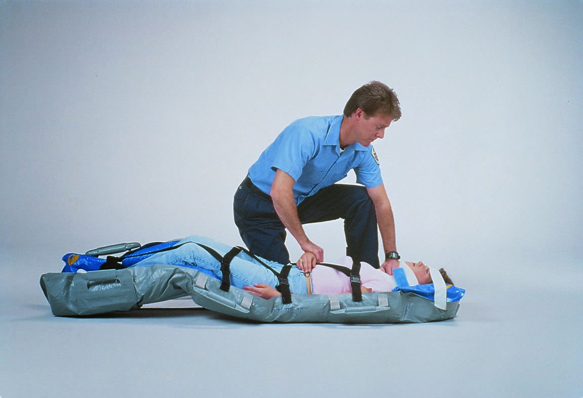
Vacuum spine boards
In an attempt to provide a more comfortable method for immobilizing the spine for transport to the emergency department, many EMS agencies are using a vacuum type splint device. Vacuum splints are flexible, waterproof shells filled with polystyrene beads similar to a very large beanbag.
Early in the application process, the splint is soft and malleable. Rescuers can push the beads into the recesses and voids between a patient and the splint. However after withdrawing the air, the splint conforms to the shape of the patient and becomes rigid.
In a side by side comparison with a rigid backboard, the vacuum splint device was judged more comfortable by the patient, was faster for rescuers to apply, and resulted in less body movement in the event the patient required tilting[15]. Healthy volunteers reported significantly less pain at 30 and 60 minutes when immobilized on a vacuum rather than a rigid spine board[16].
Pressure of about 35 mm Hg applied to the surface of the skin will significantly reduce blood flow through a capillary bed[17]. Interface pressures on good to high quality hospital mattresses vary between 30 and 60 mm Hg[18].
Researchers using pressure-mapping sensors on 20 healthy volunteers demonstrated that rigid spine boards produce the highest tissue interface pressures at the patient’s scapulae, sacrum, and heels when compared to the use of a standard, soft overlay mattress or a vacuum mattress[4].
The volunteers also rated the rigid spine board as the least comfortable device. This is consistent with a study by Lovell and Evans that demonstrated tissue interface pressures as high as 147 mm Hg with a rigid spine board, 115 mm Hg when the board was padded, but 37 mm Hg with a vacuum mattress[19].
The U.S. Army Institute of Surgical Research[20] has designated the vacuum spine board to be the immobilization method of choice during transport of patients suffering from potentially unstable thoracolumbar fractures. Unfortunately, military researchers could not demonstrate a significant reduction in pressure ulceration development associated with vacuum spine board use compared to historical controls using rigid spine boards[21].
However, more patients in the vacuum spine board group required endotracheal intubation, and thus were motionless for longer periods of time. There are anecdotal reports of awake and oriented patients becoming claustrophobic during prolonged transport while immobilized in a vacuum spine board.
Nonetheless, vacuum spine boards appear to provide a very promising alternative to the rigid spine boards historically used in EMS. These devices immobilize as well as backboards, are more comfortable for the patient, and reduce tissue interface pressures, which may reduce the development of pressure ulcerations resulting from the immobilization procedure itself.

Vacuum spine boards: Transport devices of the future


References
1. National Spinal Cord Injury Statistical Center (NSCISC). (2010). Spinal cord injury.  Facts and figures at a glance. Retrieved from https://www.nscisc.uab.edu/
2. Shafer, J. S., & Naunheim, R. S.  (2009). Cervical spine motion during extrication: A pilot study. Western Journal of Emergency Medicine, 10(2), 74-78.
3. Perry, S. D., McLellan, B., McIlroy, W. E., Maki, B. E., Schwartz, M., & Fernie, G. R.  (1999).  The efficacy of head immobilization techniques during simulated vehicle motion. Spine, 24(17), 1839–1844.
4. Keller, B. P., Lubbert, P. H., Keller, E., & Leenen, L. P. (2005). Tissue-interface pressures on three different support-surfaces for trauma patients. Injury, 36(8), 946-948.
5. March, J. A., Ausband, S. C., & Brown, L. H. (2002).  Changes in physical examination caused by use of spinal immobilization. Prehospital Emergency Care, 6(4), 421-424.
6. Bauer, D., & Kowalski, R. (1988). Effect of spinal immobilization devices on pulmonary function in the healthy nonsmoking man. Annals of Emergency Medicine, 17(9), 915-918. doi:10.1016/S0196-0644(88)80671-1
7. Schafermeyer, R. W., Ribbeck, B. M., Gaskins, J., Thomason, S., Harlan, M., & Attkisson, A. (1991). Respiratory effects of spinal immobilization in children. Annals of Emergency Medicine, 20(9), 1017-1019. doi:10.1016/S0196-0644(05)82983-X
8. Vickery, D. (2001). The use of the spinal board after the pre-hospital phase of trauma management. Emergency Medicine Journal, 18(1), 51-54. doi:10.1136/emj.18.1.51
9. Linares, H. A., Mawson,  A. R., Suarez, E., & Biundo, J. J. (1987). Association between pressure sores and immobilization in the immediate postinjury period. Orthopedics, 10(4), 571–573.
10. Mawson, A. R., Biundo, J. J. Jr., Neville, P., Linares, H. A., Winchester, Y., & Lopez, A. (1988).  Risk factors for early occurring pressure ulcers following spinal cord injury. American Journal of Physical Medicine and Rehabilitation, 67(3), 123-127.
11. Baldwin, K. M., & Ziegler, S. M. (1998). Pressure ulcer risk following critical traumatic injury. Advances in Wound Care, 11(4), 168-173.
12. Watts, D., Abrahams, E., MacMillan, C., Sanat, J., Silver, R., Van Gorder, S., Waller, M., & York, D. (1998). Insult after injury: Pressure ulcers in trauma patients. Orthopaedic Nursing, 17(40), 84-91.
13. Berg, G., Nyberg, S., Harrison, P., Baumchen, J., Gurss, E., & Hennes, E. (2010). Near-infrared spectroscopy measurement of sacral tissue oxygen saturation in healthy volunteers immobilized on rigid spine boards. Prehospital Emergency Care, 14(4), 419-424. doi:10.3109/10903127.2010.493988
14. Cooney, D. R., Wallus, H., Asaly, M., & Wojcik, S. (2013). Backboard time for patients receiving spinal immobilization by emergency medical services.  International Journal of Emergency Medicine, 6(1), 17. doi:10.1186/1865-1380-6-17
15. Johnson, D. R., Hauswald, M., & Stockhoff, C. (1996). Comparison of a vacuum splint device to a rigid backboard for spinal immobilization. American Journal of Emergency Medicine, 14(4), 369-372. doi:10.1016/S0735-6757(96)90051-0
16. Cross, D. A., & Baskerville, J. (2001). Comparison of perceived pain with different immobilization techniques. Prehospital Emergency Care, 5(3), 270-274.
17. Ryan, D. W., Allen, V., & Murray, A. (1997). An investigation of interface pressures in low air loss beds. International Journal of Clinical Practice, 51(5), 296-298.
18. Defloor, T. (2000). The effect of position and mattress on interface pressure. Applied Nursing Research, 13(1), 2-11. doi:10.1016/S0897-1897(00)80013-0
19. Lovell, M. E., & Evans, J. H. (1994). A comparison of the spinal board and the vacuum stretcher, spinal stability and interface pressure. Injury, 25(3), 179-180. doi:10.1016/0020-1383(94)90158-9
20. United States Army Institute of Surgical Research. (2012). Joint theater trauma system clinical practice guideline: Spine injury surgical management and transport. Retrieved from http://www.usaisr.amedd.army.mil/assets/cpgs/Cervical_and_Thoracolumbar_Spine_Injury_9_Mar_12.pdf
21. Mok, J. M., Jackson, K. L., Fang, R., & Freedman, B. A. (2013). Effect of vacuum spine board immobilization on incidence of pressure ulcers during evacuation of military casualties from theater. Spine Journal, 13(12), 1801-1808. doi:10.1016/j.spinee.2013.05.028

About the author
Kenny Navarro is an Assistant Professor in the Emergency Medicine Education Department at the University of Texas Southwestern Medical Center at Dallas. He coordinates all continuing education activities and assists in medical oversight for BioTel, a multi-jurisdictional EMS system composed of 14 fire/rescue agencies and more than 1,500 paramedics. Mr. Navarro serves as a Content Consultant for the AHA ACLS Project Team for Guidelines 2010 and served on two education subcommittees for NIH-funded research projects, as the Coordinator for the National EMS Education Standards Project, and as an expert writer for the National EMS Education Standards Implementation Team. Send correspondence concerning any articles in this section to Kenneth W. Navarro, The University of Texas Southwestern Medical School at Dallas, 6300 Harry Hines Blvd, MC 9134, Dallas, Texas 75390-9134, or e-mailkenny.navarro@ems1.com.

jueves, 29 de junio de 2017

Online Course Advanced Air Crew Medical by EMERGENCY EDUCATIONAL TRAINING INSTITUTE


Advanced Air Crew Medical by EMERGENCY EDUCATIONAL  TRAINING INSTITUTE 

by Dr. Ramon Reyes, MD  


This is the Air Medical Crew Course required by Helicopter and/or Air Ambulance organizations. It is a one day course with homework to be completed and sent in for completion. This meets the National Curriculum and is Florida Bureau of EMS approved. ALL MATERIAL ARE PROVIDED AT THE CLASS. Nothing is shipped to you. Please show up on the date posted at 9am.


Link to get the course  


Advanced Air Crew Medical

domingo, 11 de junio de 2017

Veinlite EMS tm

Veinlite

Veinlite EMS

Veinlite Pedi
The new Veinlite EMS is the most affordable pocket Veinlite designed for general vein imaging access in adults and children. It has 16 dual colored LEDs and is powered by two AA batteries. The Veinlite EMS provides the best value for the money.

More Information  http://www.translitellc.com/