VISITAS RECIENTES

10769850

AUTISMO TEA PDF

AUTISMO TEA PDF
TRASTORNO ESPECTRO AUTISMO y URGENCIAS PDF

We Support The Free Share of the Medical Information

Enlaces PDF por Temas

Nota Importante

Aunque pueda contener afirmaciones, datos o apuntes procedentes de instituciones o profesionales sanitarios, la información contenida en el blog EMS Solutions International está editada y elaborada por profesionales de la salud. Recomendamos al lector que cualquier duda relacionada con la salud sea consultada con un profesional del ámbito sanitario. by Dr. Ramon REYES, MD

Niveles de Alerta Antiterrorista en España. Nivel Actual 4 de 5.

Niveles de Alerta Antiterrorista en España. Nivel Actual 4 de 5.
Fuente Ministerio de Interior de España
Mostrando entradas con la etiqueta PREHOSPITAL. Mostrar todas las entradas

domingo, 13 de abril de 2025

Time for Change in Prehospital Spinal Immobilization,Suggests a Research

Time for Change in Prehospital Spinal Immobilization,Suggests a Research 
Articulo obsoleto 
Te recomiendo leer en el enlace 
http://emssolutionsint.blogspot.com/2016/09/es-necesario-inmovilizar-todos-los.html

Jim Morrissey, MA, EMT-P | From the March 2013 Issue | Tuesday, March 19, 2013
Prehospital spinal immobilization has long been held as the standard of care for victims of blunt or penetrating trauma who have experienced a mechanism of injury (MOI) forceful enough to possibly damage the spinal column. The majority of EMS textbooks stress that any significant MOI, regardless of signs and symptoms of spine injury, requires full-body immobilization, which is typically defined as a cervical collar being applied and the patient being secured to a backboard with head stabilizers in place.
This approach to patient immobilization has been accepted and implemented as the standard of care for decades with little scientific evidence justifying the practice.1–3 In addition, scant data shows that immobilization in the field has a positive effect on neurological outcomes in patients with blunt or penetrating trauma.1,4–6 In fact, several studies and articles show that spine immobilization may cause more harm than good in a select sub-set of trauma patients.5–7
Many experts question the current practice of prehospital spinal immobilization.1,2,4–15 There are now some guidelines, textbooks and an increasing number of EMS agencies that support a progressive, evidence-based approach in an effort to lessen unnecessary spinal immobilizations in the field.
It’s problematic to use MOI alone as the key indicator for prehospital spinal immobilization. In addition, the harmful sequelae and potential dangers of spine immobilization need to be considered in any field protocol. We need to examine appropriate spine injury assessment guidelines and algorithms that allow for the selective immobilization of injured patients.
We also should review immobilization devices and techniques that are more appropriate for patients who do require immobilization, or better termed, spinal motion restriction (SMR), by EMS providers.
Outdated Indicators?
It typically takes several years for EMS textbooks to catch up with new evidence and then additional time for the EMS instructional community to modify curricula and change current practice. For example, definitions of mechanisms that require spinal immobilization found in most EMS textbooks are outdated and problematic. Such indicators for potential spine injury as fall, damage to the vehicle, injury above the clavicle and mechanism of injury involving motion, are not particularly helpful when determining the best course of action in the field.
Especially troubling has been the lack of emphasis on the assessment of the patient before making a decision about immobilization. Historically, more emphasis has been placed on what happened to the vehicle or the best guess on how far someone may have fallen, instead of what actually happened to the person.
It isn’t the fall that causes injury; it’s the sudden stop at the end. The more sudden the stop, the more likely an injury results, especially if the kinetic energy was transmitted to the head and/or neck.
The physical condition of the patient must also be considered. A young, athletic person is able to withstand more forces than an elderly patient. So the spectrum of potential injuries is best determined through a detailed history and physical exam.
Vehicle damage has long been considered a strong indicator of potential spine injury, yet improvements in vehicular design and construction should change the way we look at vehicle damage. Vehicle technology and passenger protection is far superior to what it has been, particularly since the 70’s when EMS textbooks began advocating back boarding of patients in vehicles with significant damage.
Vehicle damage zones are now inherently built into newer vehicles, designed to absorb and dissipate the kinetic energy of a collision, and keep the passenger cabin relatively isolated and protected.16 An experienced paramedic once said, “The cake box might be crumpled, but the cake can be fine.”
Some textbooks accurately address this issue. Even as far back as 1990, the American Academy of Orthopaedic Surgeons addressed emergency medical responders in an extended-care environment, stating, “Patients with a positive mechanism of injury, without signs and symptoms, and with a normal pain response may be treated without full spine immobilization, if approved by your medical control physician.”17
Emergency medical personnel who work in extended-care, tactical, combat and wilderness environments have long realized the need to safely and accurately assess and clear patients regarding spinal injuries.18,19
New guidelines from Prehospital Trauma Life Support and the National Association of EMS Physicians have diminished the emphasis on immobilizing victims of penetrating trauma without neurologic deficits.20
In the setting of drowning, the 2010 evidence-based guidelines from the American Heart Association state that “Routine c-spine immobilization is a Class III (potentially harmful) unless clear trauma is evident in the history or exam, because it may unnecessarily delay or impede ventilations."21
Precautionary Immobilization
It isn’t surprising that the term and practice of “precautionary immobilization” has developed. It’s estimated that at least five million patients are immobilized in the prehospital environment in the U.S. each year. Most have no complaints of neck or back pain or other evidence of spine injury.3,11,12(See Photo 2.)
EMS personnel historically have neither been given the tools nor the authority to make informed decisions about objectively determining the need for prehospital spinal immobilization. This may be because the emergency medical community thought immobilization was always safe, conservative and always in the best interest of the patient. However, evidence now shows that, in some cases, spinal immobilization may not be in the patient’s best interest.1–3,7,8,10–13
Some prehospital care providers will admit that they often immobilize patients without evidence of spine injury because they want to avoid being questioned on arrival at the emergency department (ED). This dynamic can (and must) change with education and outreach.
Backboard-Based Immobilization
In addition to patient discomfort and anxiety associated with backboard-based immobilization, there are several potentially significant consequences. Standard immobilization requires the patient’s body to conform to a flat, hard surface. In addition, EMS secures a cervical collar around the patient’s neck and uses tape to secure the patient’s head to the board.
This practice often increases patient anxiety and has the potential to aggravate underlying injuries. Standard spinal immobilization techniques can also take away the patient’s ability to effectively protect their own airway thus significantly increasing the risk of aspiration.3–6,11,13
Patient vomiting, bleeding, airway drainage and swelling are common problems associated with trauma patients. Even with one EMS provider dedicated to the management of the airway and patient suction, it cannot be assumed that a suction catheter can handle the job when significant bleeding and/or vomiting is presented.
The continued spinal stability of a patient who is turned on their side to facilitate airway drainage and control is also questionable. Patients typically experience a significant shift in body weight and distribution, causing more movement to the spine than the immobilization process was intended to prevent.
In a comprehensive review published in Prehospital and Disaster Medicine, healthy volunteers who were immobilized on a backboard were found to be “significantly more likely to complain of pain when compared with immobilization on a vacuum mattress.” Adverse effects of backboard-based immobilization documented in this study include increased ventilatory effort, pain and discomfort.
In addition to pressure injury, the backboard may also be the cause of pain—even in otherwise healthy volunteers. The resultant posterior surface/back pain of immobilized patients has been documented to result in unnecessary radiographs and potential clinical ambiguity regarding the cause of the pain.3,22 There’s an increased cost associated with some of these complications.
It has been documented that supine patient immobilization results in a 15–20% reduction in respiratory capacity, and that respiratory effectiveness is markedly reduced by the strapping systems typically used.3,9,13 Patients are often either strapped securely, thus having diminished respiratory capacity, or loosely secured, facilitating easier breathing. Neither scenario is ideal.
The challenge is exacerbated in obese patients, the elderly and patients with such underlying diseases as congestive heart failure, COPD, asthma and pneumonia.
Done properly, immobilization in the field takes time and multiple personnel. Time delay to the ED or trauma center arrival has been cited as a significant problem for critical trauma victims. Several studies have looked at the risk vs. benefit of prehospital immobilization, with several authors and researchers questioning the value of current practices.1,2,7,8,11,15
Studies have also shown limited or no benefit of prehospital immobilization of penetrating trauma patients. (See photos on pages 32 and 33.) Unnecessary immobilization of this subset of trauma patients can result in prolonged on-scene time and delayed transport to definitive care, which may increase morbidity and mortality.4–6,14,18,23–25
Several studies show that cervical collars by themselves aren’t without risk or significant consequences.4,26–28 One study concludes that cervical collars frequently increase intracranial pressure and may be particularly harmful if used on head-injured patients.26
Another researcher observed that cervical collars “can result in abnormal distraction within the upper cervical spine in the presence of severe injury.”28 In addition, cervical collars hide areas of the head and neck, resulting in the increased possibility of missing injuries or evolving problems, such as swelling, hematoma and tracheal deviation.27,28
In addition, the longer a patient is immobilized, the more likely that cutaneous pressure ulcers will develop, most notably in the occipital, sacral or heel areas.9,12,22,29,30 This is especially true in elderly, unconscious and neurologically impaired patients.
This problem may be significantly reduced with padding or use of a vacuum mattress. Unfortunately, the vast majority of the patients who are immobilized don’t get padding in voids or areas of significant body weight/pressure or a vacuum mattress that distributes beads/padding in voids and uneven body surface areas.
The Penetrating Trauma Patient
As referenced earlier, there is a growing body of evidence that suggests penetrating trauma victims shouldn’t be routinely immobilized. Immobilization has been associated with higher mortality in patients with penetrating trauma.4–6,14,23–25
Independent studies show that whether the penetrating trauma is to the head, neck or torso, immobilization is unnecessary, interferes with and delays emergent care, and should be seriously reconsidered as the standard of care.4–6,14,23
Journal of Trauma article concluded, “Indirect spinal injury does not occur in patients with gunshot wounds to the head.” The authors state, “Protocols mandating cervical spine immobilization after a gunshot wound to the head are unnecessary and may complicate airway management.”14
Another retrospective study showed similar concerns about the use of a cervical collar with patients who have penetrating injuries to the neck. This study suggests that avoiding the collar should be the rule, and that a provider who chooses to apply a cervical collar should have good justification. The authors also suggest that frequent examination of the underlying structures and tissue is warranted if a cervical collar is used.4
A comprehensive retrospective analysis of gunshot injuries to the torso found that immobilization was of little or no benefit, even if an unstable spine fracture was present. The authors argue that airway management, including intubation, is far more complicated and problematic with prehospital spinal immobilization in place.5,6
In fact, failed airway management was reported to be the second-leading error preceding death of trauma patients, accounting for 16% of mortality in one study. This study also highlights the potential delay to definitive surgical treatment and the lack of neurologic improvement after gunshot injury to the spinal cord, suggesting that prehospital spinal immobilization is unjustified.5,6
Proper Spine Injury Assessment
For many trauma patients, a vetted field assessment criterion that focuses on the assessment of the patient rather than the mechanism of injury would obviate unwarranted immobilization.3,11,31
Many emergency medicine specialists believe an accurate, reliable, simple-to-perform spinal injury assessment could reduce spine immobilizations drastically. Thankfully, there is a trend in this direction across the nation.
The idea of “clearing” a patient of spinal injury in the field has been, and continues to be debated. However, there are prehospital spine assessment protocols that safely and accurately allow EMTs and paramedics to omit prehospital spinal immobilization in certain patients.
Some EMS experts prefer the term “selective immobilization” to “clearing” the c-spine, but the end result is the same. The end result is the reduction of the incidence of unwarranted  spinal immobilizations.
For example, the Maine spine injury assessment guidelines, developed by Peter Goth, MD, in the 1990s, have been shown to be accurate and safe.10,31,32 Several states and EMS systems around the nation use this, or a similar protocol, to help decrease the number of trauma patients being subjected to prehospital spinal immobilization.
The origin of this type of spinal assessment was initially intended to help ED physicians clinically decide if they can safely clear patients from prehospital spinal immobilization and reduce or eliminate unnecessary radiographic studies. It has been shown that the proper clinical exam and history is more accurate at predicting spine injuries than X-ray review.10,32–35
The spine injury assessment guidelines that have been adopted by multiple prehospital systems are based on the Canadian C-spine rule and the National Emergency X-Radiography Utilization Study (NEXUS) low-risk criteria. Each has similar parameters, requiring that the patient be awake, alert, conversant and without significant distracting injury or intoxication.
In addition, the guidelines further state that the physical exam should reveal no pain or tenderness to the posterior neck and back and the neurologic exam must find normal motor and sensory function in the extremities.10,18,31,33–35
Studies show that prehospital care providers can safely apply spine injury assessment criteria and not miss any clinically
significant spine injuries.10,31,32 Although these guidelines are available, training and practice is needed to become proficient at using these criteria.
Alameda County (Calif.) EMS has revised its spine injury assessment protocol to accurately reflect the current literature and research. (See Figure 1, p. 38). Its goals in 2012 were to reduce unnecessary immobilization, and use treatment modalities in the best interest of and provide the most comfort to the patient. In some cases, this meant forgoing prehospital spinal immobilization to expedite transport to a trauma center.
However, long-established norms are hard to break, and extensive training was required to make this new policy successful. EMS schools, fire departments and other EMS providers, as well as emergency department staff, needed to be exposed to the literature and trained in the new protocol.
Initial training and outreach has been well received and the early indicators have shown a significant reduction in spine immobilizations. The end result is:
>> A better understanding of the need for expeditious care under specific circumstances, in particular, the need to move rapidly when penetrating trauma is present;
>> All involved are empowered to break the paradigm of “board them all” as a result of understanding the importance of proper spinal/neurological assessment and assessment parameters that allow crews to assess for serious spinal indications and perform selective immobilization. We did the same process decades ago when we adopted rapid removal techniques for patients in lieu of spending precious minutes placing splints and half backboards on critical patients. Little or no untoward results occurred with that change in procedure;
>> More attention to patient comfort and pain instead of routine placement of trauma patients on a hard, uncomfortable platform that often put them in anatomically-incorrect positions for extended time periods, made patients unnecessarily claustrophobic lying supine and immobile and exacerbation of respiratory distress in patients due to the supine position, strap placement, and existing conditions such as CHF, COPD or morbid obesity; and
>> The ability to deploy and maximize the usage of alternative immobilization and transfer devices and stretchers such as vacuum mattresses, scoop or CombiCarriers and flexible stretchers such as Ferno and SKED stretchers and others that feature lateral patient support slats and multiple handles for convenient movement and transfer of patients. Many of these devices are better suited to patient movement in tight spaces and crew body mechanics when carrying and transferring patients down stairways and other difficult environments.
Of course, crews have to take special caution when dealing with and managing high-risk patients, including pediatric patients, the elderly and those with such degenerative bone disorders as osteoporosis. Field personnel need to be conservative while evaluating these patients and should provide spinal motion restriction when in doubt.33,34
Unconventional Options
Even with appropriate application of spine injury assessment guidelines, some patients still require some degree of prehospital spinal motion restriction. Vacuum mattresses and other break-away and flexible stretchers have been used successfully throughout Europe for years. They score well in several critical areas, including patient comfort, secure immobilization, insulation, lack of pressure sore development and, in the case of some vacuum device configurations, allow crews to utilize them without a cervical collar.12,29,30
When considering adding vacuum mattresses, vacuum stretchers or other immobilization devices to your arsenal, keep in mind that they don’t require more effort or training than using backboards. Vacuum mattresses can also effectively pad voids, distribute weight evenly and immobilize patients on their side because the device can be “molded” around the patient to best package them safely. (See photos on page 36.)
However, keep in mind that backboards still have a place, especially to restrain or slide a patient out of an extrication mess. There is also nothing that precludes you from utilizing a combination of devices such as a backboard or scoop-type stretcher to remove a patient and transfer them to a more moldable or comfortable secure surface such as a vacuum mattresses. Many systems use this combination or deploy vacuum mattresses in conjunction with flexible stretchers. (See photo, top of page 36.)
Another emerging school of thought questions the need for traditional prehospital spinal immobilization at all—even for patients who have positive evidence of a spinal column or spinal cord injury. One group of researchers who compared various extrication tools and methods found that allowing a patient to self-extricate from a vehicle with a cervical collar alone caused less movement of the spine than the use of cervical collar, KED extrication device and standard extrication techniques.36 This triggers a series of questions that are beyond the scope of this article. Groups such as the National Association of EMS Physicians and the U.S. Metropolitan Municipalities Medical Directors and Global Affiliates Consortium are carefully discussing these options and revisions to our traditional approaches to neck and spine immobilization
Conclusion
It’s appropriate for emergency personnel to immobilize certain trauma patients. However, many other trauma patients are unnecessarily immobilized by EMS. Spinal immobilization isn’t always a benign intervention. It can result in increased scene time, delay of delivery to definitive care, problematic airway management, increased patient pain or dyspnea, and unnecessary radiographic testing.
Many trauma patients can be safely and accurately assessed and treated without immobilization if they meet all criteria in prehospital spinal assessment guidelines. Extensive initial training and ongoing review is necessary for an effective selective immobilization protocol.
Science, research and multiple validated articles have changed the way EMS practices. If good patient care is the goal, it’s time that prehospital spinal immobilization be critically examined.
You could like to read 
References
1. Hauswald M, Ong G, Tandberg D, et al. Out-of-hospital spinal immobilization: Its effect on neurologic injury. Acad Emerg Med. 1998;5(3):214–219.
2. Baez AA, Schiebel N. Evidence-based emergency medicine/systematic review abstract. Is routine spinal immobilization an effective intervention for trauma patients? Ann Emerg Med. 2006;47(1):110–112.
3. Kwan I, Bunn F. Effects of prehospital spinal immobilization: a systematic review of randomized trials on healthy subjects. Prehosp Disaster Med. 2005;20(1):47–53.
4. Barkana Y, Stein M, Scope A, et al. Prehospital stabilization of the cervical spine for penetrating injuries of the neck: Is it necessary? Injury. 2000;31(5):305–309.
5. Haut ER, Kalish BT, Efron DT, et al. Spine immobilization in penetrating trauma: More harm than good? J Trauma. 2010;68(1):115–120; discussion 120–121.
6. Brown JB, Bankey PE, Sangosanya AT, et al. Prehospital spinal immobilization does not appear to be beneficial and may complicate care following gunshot injury to the torso. J Trauma. 2009;67(4):774–778.
7. Smith JP, Bodai BI, Hill AS, et al. Prehospital stabilization of critically injured patients: A failed concept. J Trauma. 1985;25(1):65–70.
8. Seamon MJ, Fisher CA, Gaughan J, et al. Prehospital procedures before emergency department thoracotomy: ‘Scoop and run’ saves lives. J Trauma. 2007;63(1):113–120.
9. Chan D, Goldberg R, Tascone A, et al. The effect of spinal immobilization on healthy volunteers. Ann Emerg Med. 1994;23(1):48–51.
10. Domeier RM, Frederiksen SM, Welch K. Prospective performance assessment of an out-of-hospital protocol for selective spine immobilization using clinical spine clearance criteria. Ann Emerg Med. 2005;46(2):123–131.
11. Kwan I, Burns F. Spinal immobilization for trauma patients (Cochrane Review). Cochrane Review; 2009; 11 http://summaries.cochrane.org/CD002803/spinal-immobilisation-for-trauma-....
12. McHugh TP, Taylor JP. Unnecessary out-of-hospital use of full spinal immobilization. Acad Emerg Med. 1998;5(3):278–280.
13. Totten VY, Sugarman DB. Respiratory effects of spinal immobilization. Prehosp Emerg Care. 1999;3(4):347–352.
14. Kaups KL, Davis JW. Patients with gunshot wounds to the head do not require cervical spine immobilization and evaluation. J Trauma. 1998;44(5):865–867.
15. Hauswald M. A re-conceptualisation of acute spinal care. Emerg Med J. Sept. 8, 2012. [Epub ahead of print].
16. Centers for Disease Control and Prevention (Sept. 6, 2012). Guidelines for Field Triage of Injured Patients. 2011; Retrieved from www.cdc.gov/Fieldtriage. Accessed Sept. 24, 2012, 2012.
17. Worsing R. Basic Rescue and Emergency Care. First Edition. Ed: American Academy of Orthopaedic Surgeons, Park Ridge, IL; 1990; 253 .
18. Goth P. Spine Injury, Clinical Criteria for Assessment and Management. Augusta, ME: Medical Care Development Publishing; 1994.
19. Morrissey J. Field Guide of Wilderness Medicine and Rescue. Third Edition Ed: Wilderness Medical Associates, Portland, ME; 2000; 30-33.
20. Stuke LE, Pons PT, Guy JS, et al. Prehospital spine immobilization for penetrating trauma: Review and recommendations from the Prehospital Trauma Life Support Executive Committee. J Trauma. 2011;71(3):763–769; discussion 769–770.
21. Berg RA, Hemphill R, Abella BS, et al. Part 5: Adult basic life support: 2010 American Heart Association Guidelines for Cardiopulmonary Resuscitation and Emergency Cardiovascular Care. Circulation. 2010;122(18 Suppl 3):S685–S705.
22. March JA, Ausband SC, Brown LH. Changes in physical examination caused by use of spinal immobilization. Prehosp Emerg Care. 2002;6(4):421–424.
23. Kennedy FR, Gonzalez P, Beitler A, et al. Incidence of cervical spine injury in patients with gunshot wounds to the head. South Med J. 1994;87(6):621–623.
24. Chong CL, Ware DN, Harris JH, Jr. Is cervical spine imaging indicated in gunshot wounds to the cranium? J Trauma. 1998;44(3):501–502.
25. Arishita GI, Vayer JS, Bellamy RF. Cervical spine immobilization of penetrating neck wounds in a hostile environment. J Trauma. 1989;29(3):332–337.
26. Davies G, Deakin C, Wilson A. The effect of a rigid collar on intracranial pressure. Injury. 1996;27(9):647–649.
27. Kolb JC, Summers RL, Galli RL. Cervical collar-induced changes in intracranial pressure. Am J Emerg Med. 1999;17(2):135–137.
28. Ben-Galim P, Dreiangel N, Mattox KL, et al. Extrication collars can result in abnormal separation between vertebrae in the presence of a dissociative injury. J Trauma. 2010;69(2):447–450.
29. Cordell WH, Hollingsworth JC, Olinger ML, et al. Pain and tissue-interface pressures during spine-board immobilization. Ann Emerg Med. 1995;26(1):31–36.
30. Luscombe MD, Williams JL. Comparison of a long spinal board and vacuum mattress for spinal immobilisation. Emerg Med J. 2003;20(5):476–478.
31. Muhr MD, Seabrook DL, Wittwer LK. Paramedic use of a spinal injury clearance algorithm reduces spinal immobilization in the out-of-hospital setting. Prehosp Emerg Care. 1999;3(1):1–6.
32. Domeier RM, Evans RW, Swor RA, et al. The reliability of prehospital clinical evaluation for potential spinal injury is not affected by the mechanism of injury. Prehosp Emerg Care. 1999;3(4):332–337.
33. Stroh G, Braude D. Can an out-of-hospital cervical spine clearance protocol identify all patients with injuries? An argument for selective immobilization. Ann Emerg Med. 2001;37(6):609–615.
34. Barry TB, McNamara RM. Clinical decision rules and cervical spine injury in an elderly patient: a word of caution. J Emerg Med. 2005;29(4):433–436.
35. Burton JH, Dunn MG, Harmon NR, et al. A statewide, prehospital emergency medical service selective patient spine immobilization protocol. J Trauma. 2006;61(1):161–167.
36. Shafer JS, Naunheim RS. Cervical spine motion during extrication: a pilot study. West J Emerg Med. 2009;10(2):74–78.


domingo, 23 de marzo de 2025

Anafilaxia (Reacciones alérgicas severas) / SHOCK ANAFILACTICO. VIDEOS

ALERGIAS, ANAFILAXIA, ASMA

"Recordar que en las nuevas guías de primeros auxilios en caso de Alergias Severas solo se podrá administrar la primera dosis de adrenalina (1-10000), para la 2ª dosis solo podrá ser administrada por un equipo medico entrenado".


by Dr.Ramón Reyes,MD


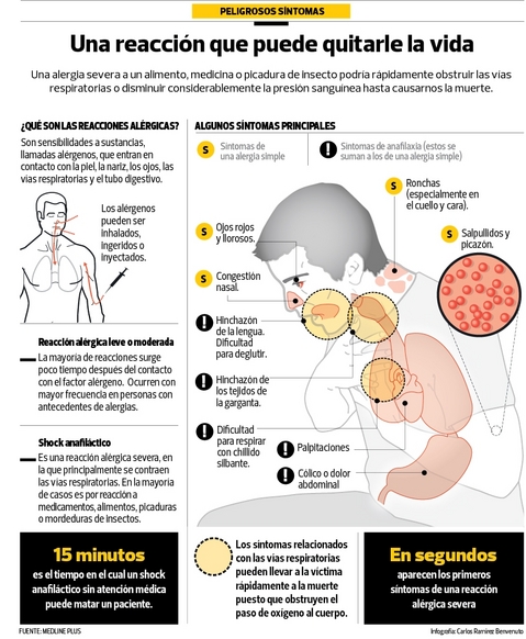
La anafilaxia es un trastorno en el cual se produce una liberación rápida de mediadores químicos con acciones biológicas potentes derivadas de células inflamatorias como los mastocitos y basófilos que provocan síntomas cutáneos, respiratorios, cardiovasculares y gastrointestinales.
Las causas más frecuentes de anafilaxia en niños difieren dependiendo de que la población estudiada represente pacientes ingresados en un servicio hospitalario o ambulatorios. La anafilaxia que se produce en el hospital se debe sobre todo a reacciones alérgicas a medicamentos y al látex, mientras que la alergia alimentaria es la causa más común de anafilaxia fuera del hospital y supone alrededor de la mitad de las reacciones anafilácticas publicadas en estudios pediátricos en los Estados Unidos, Italia y sur de Australia.
La alergia al cacahuete se ha convertido en la principal causa de anafilaxia inducida por alimentos en los países “occidentalizados” , y es responsable de la mayoría de las reacciones mortales y casi mortales.
Dentro del ámbito hospitalario el látex es un problema particular en niños sometidos a múltiples intervenciones quirúrgicas, como por ejemplo los que tienen espina bífida y trastornos urológicos, y ha llevado a muchos hospitales a emplear productos sin látex. Los pacientes con alergia al látex también pueden experimentar reacciones alérgicas a alimentos por proteínas homólogas presentes en frutas como la banana, el kiwi, la nuez, y la frutilla.


ANAFILAXIA - Guías mundiales sobre manejo de anafilaxia

La anafilaxia se define como una reacción de hipersensibilidad sistémica o generalizada, con riesgo vital. Resumimos las guías de manejo de la anafilaxia de la Organización Mundial de Alergias.

Los factores que aumentan el riesgo de una anafilaxia severa o fatal son el asma (u otra enfermedad respiratoria crónica), las enfermedades cardiovasculares, la mastocitosis, las enfermedad a tópicas severas (como la rinitis alérgica), el uso de betabloqueadores e inhibidores de la enzima convertidora de angiotensina.


Los gatillantes varían según la edad. Los alimentos son los más comunes en los niños y adultos jóvenes. Las picaduras y los medicamentos son frecuentes en adultos de edad media y mayores. Virtualmente cualquier molécula puede producir una reacción alérgica.
El diagnóstico se basa principalmente clínico (historia y exámen físico). La historia típica es la aparición súbita de síntomas y signos característicos, luego de minutos u horas de exponerse a un potencial gatillante. Los síntomas y signos progresan en pocas horas; los más frecuentes son:
  • Picazón y enrojecimiento de la piel
  • Edema y enrojecimiento de mucosas (labios, lengua, conjuntiva, etc.)
  • Alteraciones respiratorias: disnea, estridor, tos, estridor e hipoxemia
  • Hipotensión
  • Alteraciones gastrointestinales: dolor cólico y vómitos
El manejo sistemático y rápido es esencial; es aplicable en cualquier momento de la crisis aguda, con cualquier gatillante y puede ser realizado por cualquier profesional médico con entrenamiento básico. El protocolo de manejo debe estar escrito, visible y constantemente revisado para que sea de utilidad. Las pruebas de laboratorio pueden ayudar a precisar el diagnóstico después de la emergencia. Se puede medir niveles de triptasa, con una muestra obtenida 15 minutos a 3 horas después del inicio del cuadro, siendo lo más recomendable la medición seriada. La triptasa generalmente está normal en anaflixia por alimentos y sin hipotensión. Los niveles de histamina se pueden obtener 15 minutos a 1 hora después del inicio, pero se requiere mantener la muestra en condiciones extremadamente complejas.
  1. Elimine el gatillante (por ej. suspender un medicamento)
  2. Evalúe vía aérea, ventilación, circulación, estado mental, piel y peso del paciente.
  3. Pida ayuda
  4. Administre adrenalina intramuscular en el tercio medio de la cara anterolateral del muslo (0,01 mg/kg, máximo 0,5 mg en adultos y 0,3 mg en niños). Repita cada 5-15 minutos según necesidad.
  5. Posiciones al paciente sobre su espalda y levante las extremidades inferiores
  6. Evalúe utilizar oxígeno suplementarios, fluidos endovenosos (solución fisiológica; 1-2 litros en adultos o 10 ml/kg en niños) y/o reanimación cardiopulmonar, según el caso.
  7. Monitorice los signos signos vitales continuamente.
La adrenalina se utiliza como un medicamento esencial para tratar la anafliaxia, debido a sus efectos alfa-1 (vasoconstricción sintética, que produce disminución del edema de mucosas y aumenta la presión arterial), beta-1 (efecto inótropo y cronótropo) y beta-2 (broncodilatador). La administración de adrenalina es más importante que el uso de antihistamínicos y corticoides en la etapa precoz de la anafilaxia. Se debe administrar intramuscular, en el tercio medio de la cara anterolateral del muslo, tan pronto se diagnostique o sospeche fuertemente una anafilaxia, en dosis de 0,01 mg/kg (concentración de 1 mg/ml) hasta un máximo de 0,5 mg en adultos y 0,3 mg en niños. Se puede repetir la dosis cada 5-15 minutos. El retraso en la administración de adrenalina se ha asociado a mortalidad, encefalopatía hipóxico-isquémica y anafilaxia bifásica (recurrencia en 8-10 horas).
Si el paciente ya se encuentra en shock, la adrenalina se debe administrar como infusión continua intravenosa, titulando según la presión arterial y la frecuencia cardiaca. Si el paciente está en paro cardiorrespiratorio, se debe administrar adrenalina en bolos intravenosos.
Con las dosis recomendadas, se puede producir palidez, temblor, palpitaciones y cefalea. Con dosis mayores o administración intravenosa se pueden producir arritmias ventriculares, crisis hipertensas o edema pulmonar. La adrenalina no está contraindicada en pacientes con patología cardiovascular.
No están claras las recomendaciones respecto a los medicamentos de segunda línea. Las guías mundiales dicen que no está claro el rol de ninguno de ellos y son secundarion con respecto a la adrenalina.
  • Antihistamínicos H1: disminuyen el prurito, enrojecimiento, angioedema, los síntomas nasales y los síntomas oculares. No sirven para tratar la obstrucción de la vía aérea, la hipotensión ni el shock.
  • Agonistas beta-2: sólo sirven para tratar la obstrucción respiratoria baja. No alivian la obstrucción respiratoria alta, la hipotensión ni el shock.
  • Glucocorticoides: inician su acción después de varias horas y sus beneficios son más teóricos que reales.
  • Antihistamínicos H2: disminuyen la cefalea y el enrojecimiento, pero pueden aumentar la hipotensión.
Los casos refractarios deben ser manejados por médicos especialistas en centros médicos de alta complejidad. Si requieren intubación, debe realizarla el operador más capacitado por su alta complejidad, con una adecuada preoxigenación (3-4 minutos). Algunos pacientes requieren vasopresores por infusión contínua, pero no está claro cuál de ellos es la mejor opción. En lo pacientes que utilizan betabloqueadores, se puede utilizar glucagón. Algunos pacientes pueden requerir atropina.
Hay algunas consideraciones respecto al manejo en situaciones especiales:
  • Embarazo: manejo común, salvo que se debe posicionar inclinada sobre su lado izquierdo después de las 20 semanas de embarazo y monitorizar el bienestar fetal después de las 24 semanas.
  • Niños: poner especial énfasis en una correcto cálculo de la dosis de epinefrina y sus complicaciones.
  • Adultos mayores: frecuentemente complicada por patología cardiovascular o uso de betabloqueadores. No está contraindicada la adrenalina.

Conclusiones:

  • El riesgo de anafilaxia severa aumenta en los pacientes con patología cardiovascular y respiratoria subyacente.
  • El diagnóstico es clínico y se basa en alteraciones de piel, mucosas, respiratorias, cardiovasculares y grastrointestinales.
  • El manejo debe ser sistemático y rápido. Elimine el gatillante, evalue el ABC y administre adrenalina.
  • La adrenalina es el tratamiento más importante. No está clara la utilidad de los otros medicamentos.

Artículo fuente


Enlace Guía-ABE visualizar y si quieres el pdf pincha 
aquí


Nuevas Guias RCP 2020-2025 AHA/ILCOR 


SALUD
Inmunoterapia Solución a las alergias
Jatnna Concepcíon
Quizás tienes años lidiando con la molesta alergia que no te deja llevar una vida normal y que te ata a una serie de cuidados excesivos. ahora ya no tienes barreras que te impidan vivir plenamente porque tu problema ya tiene una solución.
Las enfermedades alérgicas son la condición de tener respuestas inmunológicas “erróneas” que desatan síntomas variados ante cualquier tipo de contacto con sustancias (alergenos) que generalmente no son dañinas para personas que no sufran de estas alergias. Las alergias se pueden presentar en cualquier edad, y los afectados suelen ser personas que por herencia genética son predispuestos a desarrollar reacciones alérgicas.
La inmunoterapia es un tratamiento para las alergias que consiste en suministrar al paciente de vacunas que contienen los mismos alergenos ante los que reacciona para desensibilizarlo y que su cuerpo pueda adaptarse a ellas poco a poco hasta curarse en pocos años.
No todos los tipos de alergias se pueden tratar con inmunoterapia, en algunos casos el médico alergólogo debe tratar al paciente con farmacología y va reduciendo la exposición del paciente en cuanto a los alergenos identificados como detonantes de sus reacciones alérgicas, explica la doctora Victoria Michelén, propietaria del Centro de Inmunoterapia de Asma y Alergia Dacarette, en República Dominicana.
La inmunoterapia bien llevada suele tener una duración de 3 a 5 años, pero el paciente ya siente una mejoría en su salud desde antes de cumplir los seis meses de su tratamiento y, dependiendo de la escuela que siga el alergólogo, puede indicarse a las personas a partir de los 5 años.
Alergenos más comunes
o Los ácaros, animalitos microscópicos que se alimentan de la piel que descamamos y el cabello que se cae naturalmente y viven en la cama, la ropa guardada, etc.
o Algunos alimentos, entre ellos: la leche y sus derivados, los cítricos, los mariscos, maníes, nueces, soya, entre otros.
o Algunos medicamentos.
o Picaduras de insectos.
o Polen, lo que no es frecuente en República Dominicana por tener una estación casi única durante todo el año.
o Animales como los perros, gatos y las cucarachas.
Mitos sobre las alergias
1.  Aún muchos dicen que las alergias no tienen cura, pero esto es falso porque la inmunoterapia es un tratamiento que científicamente ofrece la solución a este mal.

2. Las vacunas que se usan en la inmunoterapia tienen cortisona, estas vacunas sólo contienen el alergeno ante el que reacciona el paciente y solución salina.

3. Las vacunas se inyectan de forma intravenosa, si se le inyecta a un paciente alérgico, una de las vacunas con el alergeno ante el que reacciona de manera intravenosa se hace mala práctica médica y se pone en riesgo la vida de éste Las inyecciones deben hacerse de forma subcutánea y nunca intravenosa en la inmunoterapia.

4. Las alergia no suelen ser causa de muerte,  pero sí pueden tener consecuencias fatales, como la anafilaxis al ser detonante de bronco espasmos, hipertensión severa, pérdida de la conciencia, cierre laríngeo y hasta ataques cardíacos en cuestiones de segundos.
Beneficios de la inmunoterapia:
o Detiene la marcha alérgica
o Elimina o disminuye de un 90 a 95% los signos, a la vez que evita que surjan nuevos síntomas.
o Evita que se afecten nuevos órganos
o Disminuye el uso de medicamentos.
o Mejora la calidad de vida del paciente.
Articulos relacionados REACCIONES ALERGICAS


posted by Dr. Ramon ReyesMD 🧩 𓃗 #DrRamonReyesMD 🧩 𓃗 @DrRamonReyesMD


Pinterest

Twitter

Blog

Gracias a todos el Canal somos mas de  1000 participantes en WhatsApp. Recordar este es un canal y sirve de enlace para entrar a los tres grupos; TACMED, TRAUMA y Científico. ahí es que se puede interactuar y publicar. Si le molestan las notificaciones, solo tiene que silenciarlas y así se beneficia de la informacion y la puede revisar cuando usted así lo disponga sin el molestoso sonido de dichas actualizaciones, Gracias a todos Dr. Ramon Reyes, MD Enlace al 


Enlace a Científico https://chat.whatsapp.com/IK9fNJbihS7AT6O4YMc3Vw en WhatsApp 

tiktok

TELEGRAM Emergencias https://t.me/+sF_-DycbQfI0YzJk  

TELEGRAM TACMED https://t.me/CIAMTO

AVISO IMPORTANTE A NUESTROS USUARIOS
Este Blog va dirigido a profesionales de la salud y público en general EMS Solutions International garantiza, en la medida en que puede hacerlo, que los contenidos recomendados y comentados en el portal, lo son por profesionales de la salud. Del mismo modo, los comentarios y valoraciones que cada elemento de información recibe por el resto de usuarios registrados –profesionales y no profesionales-, garantiza la idoneidad y pertinencia de cada contenido.

Es pues, la propia comunidad de usuarios quien certifica la fiabilidad de cada uno de los elementos de información, a través de una tarea continua de refinamiento y valoración por parte de los usuarios.

Si usted encuentra información que considera errónea, le invitamos a hacer efectivo su registro para poder avisar al resto de usuarios y contribuir a la mejora de dicha información.


El objetivo del proyecto es proporcionar información sanitaria de calidad a los individuos, de forma que dicha educación repercuta positivamente en su estado de salud y el de su entorno. De ningún modo los contenidos recomendados en EMS Solutions International están destinados a reemplazar una consulta reglada con un profesional de la salud.

Ambulancias para Pacientes Bariátricos (Obesos mórbidos). Bariatric patients "Ambulance"

bariatric patient
bariátrico, ca

Del ingl. bariatric, y este del gr. βαρύς barýs 'pesado' y ἰατρικός iatrikós 'concerniente a la medicina, medicinal, curativo'.

1. adj. Med. Perteneciente o relativo a la bariatría.

https://dle.rae.es/bari%C3%A1trico

obesidad
 

Del lat. obesĭtas, -ātis.

1. f. Cualidad de obeso.

Sin.:
  • gorduraadiposidadadiposispesadez.
Ant.:
  • escualidezdelgadez.

Sinónimos o afines de obesidad
  • gorduraadiposidadadiposispesadez.
Antónimos u opuestos de obesidad
  • escualidezdelgadez.

mórbido, da
 

Del lat. morbĭdus, y este der. de morbus 'enfermedad'.

1. adj. Que padece enfermedad o la ocasiona.

Sin.:
  • patológicoenfermizomorbosomalsanoinsanonocivopernicioso.
Ant.:
  • sanosaludable.
  • beneficiosoinocuo.

2. adj. Blandodelicadosuave.

Sin.:
  • blandodelicadosuaveflácidofofoblandengue.
Ant.:
  • duroáspero.

Sinónimos o afines de mórbido, da
  • patológicoenfermizomorbosomalsanoinsanonocivopernicioso.
  • blandodelicadosuaveflácidofofoblandengue.
Antónimos u opuestos de mórbido, da
  • sanosaludable.
  • beneficiosoinocuo.
  • duroáspero.

TQ Torniquete en paciente bariátrico "Obeso Mórbido" 


¿QUÉ ES UNA AMBULANCIA BARIÁTRICA?
Ver video 

Facebook 

Obesidad Mórbida: Una Amenaza Global y la Lucha por su Reducción

Resumen

La obesidad mórbida es una condición médica grave que afecta a millones de personas en todo el mundo, con implicaciones significativas para la salud pública y la economía global. El Dr. Ramón Reyes, conocido como DrRamonReyesMD, ha dedicado su labor en su blog, EMS Solutions International, a concienciar sobre los peligros de esta enfermedad y a promover estrategias efectivas para su prevención y tratamiento. Como ex paciente de obesidad mórbida que logró recuperarse mediante cirugía bariátrica y cambios en el estilo de vida, el Dr. Reyes ofrece una perspectiva única y valiosa en esta lucha. Este artículo analiza los riesgos asociados a la obesidad mórbida, las intervenciones disponibles y la importancia de abordar esta condición desde una perspectiva científica y realista.

Introducción

La obesidad mórbida, definida como un índice de masa corporal (IMC) superior a 40 kg/m², está asociada con un aumento significativo en la morbilidad y mortalidad. Contribuye al desarrollo de enfermedades crónicas como diabetes tipo 2, hipertensión, enfermedades cardiovasculares y ciertos tipos de cáncer. A pesar de la evidencia científica que respalda estos riesgos, algunas corrientes ideológicas contemporáneas buscan minimizar o reinterpretar la obesidad, presentándola como una variante más de la diversidad corporal y desvinculándola de sus implicaciones para la salud. El Dr. Ramón Reyes ha sido un crítico activo de estas tendencias, enfatizando la necesidad de reconocer la obesidad mórbida como la enfermedad grave que es y de implementar estrategias basadas en evidencia para su manejo.

Impacto de la Obesidad Mórbida en la Salud

La obesidad mórbida tiene múltiples efectos adversos en la salud:

Enfermedades Metabólicas: Incrementa el riesgo de desarrollar resistencia a la insulina y diabetes tipo 2.

Enfermedades Cardiovasculares: Aumenta la probabilidad de hipertensión, dislipidemia y eventos cardiovasculares como infartos e ictus.

Problemas Respiratorios: Está asociada con apnea del sueño y síndrome de hipoventilación.

Trastornos Musculoesqueléticos: Contribuye al desarrollo de osteoartritis y dolor crónico debido al exceso de peso sobre las articulaciones.

Cáncer: Se ha relacionado con un mayor riesgo de ciertos tipos de cáncer, incluyendo el de mama, colon y endometrio.


Además, la obesidad mórbida puede llevar a complicaciones adicionales como enfermedad hepática grasa no alcohólica, enfermedad renal crónica y trastornos psicológicos, incluyendo depresión y baja autoestima. Estas comorbilidades no solo afectan la calidad de vida del individuo, sino que también aumentan la carga sobre los sistemas de salud.

Costos Económicos de la Obesidad Mórbida

La carga económica de la obesidad mórbida es significativa:

Gastos Médicos Directos: Incluyen costos de hospitalización, medicamentos y tratamientos relacionados con las complicaciones de la obesidad.

Pérdida de Productividad: Días laborales perdidos, disminución de la productividad y jubilaciones anticipadas debido a discapacidades relacionadas.

Gastos Indirectos: Costos asociados con la necesidad de adaptar infraestructuras y servicios para personas con obesidad mórbida.


Según datos de la Organización para la Cooperación y el Desarrollo Económicos (OCDE), la obesidad reduce el PIB en un 3,3% en promedio en los países miembros, debido a los costos mencionados anteriormente. Además, se estima que los sistemas de salud gastan entre un 2% y un 7% de su presupuesto total en tratar enfermedades relacionadas con la obesidad.

La Experiencia del Dr. Ramón Reyes

El Dr. Ramón Reyes, a través de su blog EMS Solutions International, ha compartido su experiencia personal con la obesidad mórbida y su recuperación mediante cirugía bariátrica y cambios en el estilo de vida. Su testimonio sirve como inspiración y evidencia de que, con las intervenciones adecuadas, es posible superar esta enfermedad. El Dr. Reyes enfatiza la importancia de abordar la obesidad mórbida desde una perspectiva científica, evitando la trivialización de la enfermedad y promoviendo estrategias basadas en evidencia para su prevención y tratamiento.

Estrategias para el Manejo de la Obesidad Mórbida

El tratamiento de la obesidad mórbida requiere un enfoque multidisciplinario que incluya cambios en el estilo de vida, intervenciones farmacológicas y, en casos seleccionados, procedimientos quirúrgicos.

1. Modificaciones en el Estilo de Vida

Alimentación Saludable: Adoptar una dieta equilibrada rica en frutas, verduras, proteínas magras y granos integrales. Reducir la ingesta de azúcares añadidos, grasas saturadas y alimentos ultraprocesados.

Actividad Física Regular: Realizar al menos 150 minutos de ejercicio moderado a la semana, como caminar, nadar o practicar algún deporte.

Apoyo Psicológico: Participar en terapias conductuales para abordar patrones de alimentación desordenados y mejorar la relación con la comida.


2. Tratamiento Farmacológico

En casos donde las modificaciones en el estilo de vida no son suficientes, se pueden considerar medicamentos aprobados para el tratamiento de la obesidad:

Orlistat: Inhibe la absorción de grasas en el intestino.

Fentermina/Topiramato: Suprime el apetito y aumenta la sensación de saciedad.

Naltrexona/Bupropión: Modula el apetito y reduce los antojos.

**Liraglutida y Semaglutida:**


Una ambulancia bariátrica es una ambulancia diseñada y equipada para transportar personas con sobrepeso. Este tipo de vehículo puede estar adaptado tanto para la prestación de servicios de transporte sanitario urgente (TSU) como transporte sanitario no urgente (TSNU).
La ambulancia bariátrica, también conocida como UVI Bariátrica, es un vehículo diseñado para el traslado, de manera confortable, de pacientes con un peso superior a los 150 kilos (330 Libras)

CARACTERÍSTICAS DE LA AMBULANCIA BARIÁTRICA
Este tipo de ambulancias son más grandes que las ambulancias habituales y están equipadas con grandes cunas reforzadas con ruedas para pacientes, así como rampas que son capaces de soportar cargas pesadas.

Este tipo de vehículo disponen de un conjunto de camilla eléctrica y raíl móvil con una elevada capacidad de carga igual o superior a los 300 kg, incorporando un sistema de anclaje.

La ambulancia bariátrica no solo protege el bienestar y la dignidad de los pacientes que las necesitan, sino que también protegen a los trabajadores sanitarios, que corren el riesgo de sufrir lesiones al intentar transportar a pacientes con sobrepeso sin los equipos necesarios para ello.

Estos vehículos suelen adoptar la forma de tipo “cajón”. En general, las ambulancias bariátricas resuelven la carga del paciente una vez acomodado en la camilla, hasta el interior de la ambulancia y, en caso de tener mayores dimensiones, permiten una mayor comodidad durante el traslado y facilitan la asistencia en ruta.

¿CUÁNDO SE USAN LAS AMBULANCIAS BARIÁTRICAS?
Como ya hemos comentado, este tipo de vehículo se utiliza para realizar el transporte sanitario a pacientes con un peso superior a los 150kg.

Este tipo de ambulancia se utiliza mucho en países como Estados Unidos o Canadá, donde un porcentaje alto de la población sufre problemas de sobrepeso. En España también están presentes, pero su uso es mucho menos habitual y no está tan extendido como en lo países nombrados anteriormente.

Esta tipología de ambulancia también se emplea para el traslado de recién nacidos (neonatal) que precisen de incubadora y garantizan un trayecto con la máxima seguridad.

FLOTA DE GRUP LA PAU
En Grup La Pau contamos con una ambulancia bariátrica adaptada para el traslado de pacientes con obesidad mórbida, y está pensada para dar el mayor grado de confort posible a este tipo de personas durante el servicio. Contiene una cama articulada de 2 metros, que para su acceso y salida de la ambulancia dispone de una rampa y un cabestrante con el fin de facilitar y suavizar la subida y bajada del paciente.

Fuente


¿Qué es una camilla bariátrica de ambulancia?
25 de agosto de 2022
KARTSANA Camilla Bariátrica
Una camilla bariátrica permite el transporte de pacientes voluminosos en ambulancias y hospitales. Son camillas adaptadas y diseñadas para soportar 300 Kg de peso. Su nombre proviene del término bariatría, siendo su raíz del griego “bari” que significa “peso”.

Actualmente las camillas bariátricas de ambulancias son eléctricas en su mayoría, permitiendo al Técnico de Emergencia Sanitario subir y mover la camilla sin esfuerzos ni riesgos de sufrir lesiones transportando un paciente con sobrepeso. Asimismo, la camilla va acompañada de un raíl telescópico capaz de soportar la misma capacidad de carga, el cual va anclado en la ambulancia, adaptada también para tal fin.

Traslados de especial cuidado
Los pacientes con mayor volumen y peso o pacientes bariátricos requieren una atención especial, es importante garantizar la seguridad y la estabilidad en el traslado a la vez que su comodidad.

La camilla bariátrica puede ser una única unidad o se puede optar por adaptar un accesorio de ensanchamiento sobre camillas que soporten los 300 Kg. Las camillas bariátricas ocupan más espacio dentro de la ambulancia, por lo tanto, esta última opción resulta más flexible, ya que la camilla se puede utilizar tanto en su versión estándar como bariátrica.

La camilla bariátrica de KARTSANA
KARTSANA dispone de dos tipos de accesorios o kit bariátricos para sus camillas:

El kit bariátrico
Es un accesorio de ensanchamiento lateral que consta de un sistema que permite la instalación instantánea, anclando los ejes del kit en pestillos preinstalados en la camilla. Cuenta con 2 versiones para la camilla POWER BRAVA TG-1000 y para la serie Júpiter TG-880C2 y C4.


Kit bariátrico
Consiste en un accesorio de ensanchamiento que se coloca en la superficie del paciente mediante un sistema de guías a encajar en posicionadores que aseguran la instalación. Al igual que el sistema CLICK, este kit bariátrico tiene opciones para las camillas POWER BRAVA TG-1000 y para Júpiter TG-880C2 y C4.

obesidad 
Accesorio de ensanchamiento de la superficie del paciente para transporte de pacientes voluminosos. El sistema permite la instalación en 6 minutos, es plegable y se suministra con los cinturones, colchones laterales y una cómoda bolsa de transporte.

LEER en el enlace 
https://emssolutionsint.blogspot.com/2010/12/obesidad-infantil-un-problema-de.html
Practice professionalism with bariatric patients 

As individuals we are entitled to our opinions, but understand the causes of obesity before commenting on it
EMS News in Focus 
by Arthur Hsieh
Earlier this week, an EMS1 reader reached out to me via email, lamenting about some of the crass comments that were written about obese patients. Indeed, it's pretty interesting how some of our colleagues view obese patients. Fortunately, most of the comments on the thread were in rebuttal to the few crass ones.
Obesity is a major health issue for many in our country. According to the Centers for Disease Control nearly 36 percent of the U.S. population is obese, which is defined as having a body mass index (BMI) of 30 or more, and 6.3 percent are morbidly obese (having a BMI of 40 or more). The health issues associated with being very overweight are significant, and are a factor in the patient population EMS serves.
The reasons for being obese are many. Medical conditions, the inability to conveniently buy healthy foods, a lifestyle that promotes poor eating habits, and not making exercise a priority all contribute to the issue. Making a deliberate choice to be obese is not a factor.
As individuals we are entitled to our opinions – this is a free country, after all. But one should know the facts and better understand the causes of an issue before spouting off something that is simply embarrassing to read.
While we're on this issue, recognize that injury rates to EMS workers are significant. According to the National Institutes of Occupational Safety and Health, over 27,000 EMS provider injuries were reported in 2011. A majority of injuries occur during exertion, and center around the neck and trunk (back). It's crucial that we are in good physical shape, use excellent ergonomics and take advantage of appropriately designed and tested lifting equipment to make our job safer.
The fact is, we perform work that is abrupt in nature, often interspersed with long intervals of sitting around. It's a perfect recipe for an injury to occur.


Bariatric patients by EMS1
Read more http://goo.gl/Dj7sde


View this post on Instagram

A post shared by DR RAMON REYES MD (@drtolete) on


About the author
EMS1 Editorial Advisor Art Hsieh, MA, NREMT-P currently teaches at the Public Safety Training Center, Santa Rosa Junior College in the Emergency Care Program. Since 1982, Art has worked as a line medic and chief officer in the private, third service and fire-based EMS. He has directed both primary and EMS continuing education programs. Art is a textbook author, has presented at conferences nationwide, and continues to provide patient care at an EMS service in Northern California. Contact Art atArt.Hsieh@ems1.com.


A video training presentation designed solely for the employees of S.T.A.R. Ambulance Service on the safe and proper transportation of bariatric patients. While the presentation does attempt to best follow the guidelines set forth by the manufacturers, the statements and procedures used in this video have not been approved by nor necessarily represent those of either the Stryker or Tran Safe corporations.

Bariatric patients by EMS1 Art By Paul Combs
Read more http://goo.gl/Dj7sde

Moving bariatric patients just got easier

The latest BEAR connects to a stair chair to safely transport large patients up and down multiple stories

Expert Analysis: Earlier this week, an EMS1 reader reached out to me via email, lamenting about some of the crass comments that were written about obese patients. Indeed, it's pretty interesting how some of our colleagues view obese patients. Fortunately, most of the comments on the thread were in rebuttal to the few crass ones. Practice professionalism with bariatric patients

Obesity is a growing problem, no pun intended. The average weight for men 20 to 74 years old rose dramatically from 166.3 pounds in 1960 to 191 pounds in 2002. The new BEAR Stair Chairhelps EMS providers address this challenge.

BEAR-iatrics, Inc is known for the BEAR, or Bariatic Equalizing Abdominal Restraint, which I’vewritten about earlier. The BEAR products all share one design principle: They keep the loose mass of the obese patient from shifting around. If the abdomen isn’t moving around, it’s easier to control and move the patient.
One or two belts simply are not enough to safely secure an obese patient to a tiny seat. Frankly I’ve never used a stair chair on a very large patient for this very reason. But often there just isn’t any good way to get these patients down stairways. Where are the biggest patients when you arrive on scene? The second story, of course — unless there is a third floor. Then you will inevitably find them up there.
How it works
The latest version of the BEAR is built for use with a stair chair. It’s a great solution to a tough problem. Most stair chairs have fairly small seats, usually only about 20 inches wide. This helps make them easy to store. But with an obese patient they can be a little scary.
The BEAR attaches to the frame of the stair chair and wraps around the patient’s abdomen in two directions. The lower part then comes up and around the thighs and hips. The top bands feed through clever pockets that allow for length adjustment.
It does a great job keeping the patient and their abdominal mass centered and in place. This opens up an important new means of moving these patients down stairs.
Power combo
We now have newer powered stair chairs available with astonishing weight capacities. Used in junction with a powered chair, the BEAR Stair Chair opens up a whole new realm of possibilities.
In a video of a demonstration at the 2013 EMS World Expo, you can see a very large patient being moved both up and down stairs in the Ferno EZ Glide stair chair with Power Traxx. At one point he tries to shift his mass in the chair. This would be disastrous in most real world situations. The BEAR Stair Chair does a great job of keeping him firmly in place.
The EZ Glide makes moving him look so easy. Tom Cox of Ferno even shows off a little by briefly maneuvering the chair one-handed. It is plain to see this is a one-two combination of products that really works. For more information on the BEAR Stair Chair, check out BEAR-iatrics website.


Bariatric patients by EMS1 ART by PAul Combs
Read more http://goo.gl/Dj7sde

About the author
Dan White, EMT-P works for Intersurgical, Inc. as the National Account Manager for EMS. Immediately prior he ran Arasan, LLC. He served as Sales & Marketing Director for Truphatek, Inc. and before that Director of Corporate Planning & Product Development for AllMed. He has been certified as a paramedic since 1978 and an EMS and ACLS instructor since 1981. Dan has designed many emergency medical products since his first, the White Pulmonary Resuscitator, including the Prolite Speedboad, Cook Needle Decompression Kit and RapTag Triage System. His more recent EMS product designs are the Arasan Ultra EMS Coat and the B2 Paramedic Helmet. To contact Dan, email dan.white@ems1.com.

Related: How Britain's fattest woman - weighing 55 stone - was rescued from her flat.... with TWO cranes, SEVEN police cars, TWO fire engines and ELEVEN medics!


Read more: http://www.dailymail.co.uk/news/article-3063476/How-Britain-s-fattest-woman-rescued-flat-TWO-cranes-SEVEN-police-cars-TWO-fire-engines-ELEVEN-medics.html#ixzz3ZNPeMyIN
Follow us: @MailOnline on Twitter | DailyMail on Facebook


Free Download PDF: Task Performance and Health Improvement Recommendations for emergency medical service Practitioners. NAEMT http://goo.gl/nYhC6e

MANUAL DE ATENCIÓN AL PARTO EN EL ÁMBITO EXTRAHOSPITALARIO. Ministerio de Sanidad, Servicios Sociales e Igualdad. España


¿Qué es el parto velado "Parto Empelicado" o nacer con bolsa intacta? by NATALBEN.com


Balística de las heridas: introducción para los profesionales de la salud, del derecho, de las ciencias forenses, de las fuerzas armadas y de las fuerzas encargadas de hacer cumplir la ley http://emssolutionsint.blogspot.com/2017/04/balistica-de-las-heridas-introduccion.html
Guía para el manejo médico-quirúrgico de heridos en situación de conflicto armado by CICR http://emssolutionsint.blogspot.com/2017/09/guia-para-el-manejo-medico-quirurgico.html


CIRUGÍA DE GUERRA TRABAJAR CON RECURSOS LIMITADOS EN CONFLICTOS ARMADOS Y OTRAS SITUACIONES DE VIOLENCIA VOLUMEN 1 C. Giannou M. Baldan CICR http://emssolutionsint.blogspot.com.es/2013/01/cirugia-de-guerra-trabajar-con-recursos.html


Manual Suturas, Ligaduras, Nudos y Drenajes. Hospital Donostia, Pais Vasco. España http://emssolutionsint.blogspot.com/2017/09/manual-suturas-ligaduras-nudos-y.html


Técnicas de Suturas para Enfermería ASEPEYO y 7 tipos de suturas que tienen que conocer estudiantes de medicina http://emssolutionsint.blogspot.com/2015/01/tecnicas-de-suturas-para-enfermeria.html


Manual Práctico de Cirugía Menor. Grupo de Cirugia Menor y Dermatologia. Societat Valenciana de Medicina Familiar i Comunitaria http://emssolutionsint.blogspot.com/2013/09/manual-practico-de-cirugia-menor.html

Protocolo de Atencion para Cirugia. Ministerio de Salud Publica Rep. Dominicana. Marzo 2016 http://emssolutionsint.blogspot.com/2016/09/protocolo-de-atencion-para-cirugia.html
Manual de esterilización para centros de salud. Organización Panamericana de la Salud http://emssolutionsint.blogspot.com/2016/07/manual-de-esterilizacion-para-centros.html

Bariatric patients "Ambulance" by EMS1

http://emssolutionsint.blogspot.com/2017/01/bariatric-patients-by-ems1.html


Fisiopatología del Transporte Sanitario Terrestre

Clases "Tipos" de Ambulancias en España (Union Europea). UNE-EN 1789:2007 "Real Decreto 836/2012 del 25 de mayo"

Clases "Tipos" de Ambulancias en España (Union Europea). UNE-EN 1789:2007 "Real Decreto 836/2012 del 25 de mayo"

posted by Dr. Ramon Reyes, MD 🧩  #DrRamonReyesMD 🧩 𓃗 @DrRamonReyesMD


Descubrí los siguientes consejos básicos para prevenir o revertir el sobrepeso y la obesidad Leer http://emssolutionsint.blogspot.com/2023/03/obesidad-infografia-by-msp.html y además ¿Qué es la inclusión forzada? "corrección política" y la ideología progresista o "woke" https://emssolutionsint.blogspot.com/2024/08/que-es-la-inclusion-forzada-correccion.html


‼️Un vistazo al pasado: Daniel Lambert‼️
En un momento dado, Daniel Lambert fue considerado la persona más pesada de la historia de la humanidad. A los 18 años, Lambert comenzó a trabajar como guardia de prisión y era muy respetado por su capacidad para hacerse amigo de muchos prisioneros y ayudarlos a rehabilitarse.
En una ocasión caminó 11 kilómetros desde Woolwich hasta la City de Londres "con mucha menos fatiga aparente que varios hombres de tamaño mediano que formaban parte del grupo". En términos de fuerza, Lambert podía cargar fácilmente 254 kg y tenía la capacidad de mantenerse en pie sobre una pierna.
En 1805, Lambert decidió exhibirse y cobrar un chelín a quienes lo visitaran en su casa del número 53 de Piccadilly. Trabajaba cinco horas al día y hablaba con casi 400 visitantes diarios sobre diversos temas relacionados con los perros y los deportes.
Lambert también promulgó una norma según la cual todos los que entraban en su casa debían quitarse el sombrero. Cuando un visitante se negó a quitárselo "incluso si el rey estaba presente", Lambert respondió amablemente: "Entonces, por Dios, señor, debe abandonar esta habitación de inmediato, ya que no lo considero una muestra de respeto hacia mí, sino hacia las damas y caballeros que me honran con su compañía".
Lambert falleció repentinamente en 1809 a la edad de 39 años. Su ataúd era tan grande que tuvieron que tirar de él 20 hombres. En el momento de su muerte, pesaba 335 kg (739 lb).


Pinterest

Twitter

Blog

Gracias a todos el Canal somos mas de  1000 participantes en WhatsApp. Recordar este es un canal y sirve de enlace para entrar a los tres grupos; TACMED, TRAUMA y Científico. ahí es que se puede interactuar y publicar. Si le molestan las notificaciones, solo tiene que silenciarlas y así se beneficia de la informacion y la puede revisar cuando usted así lo disponga sin el molestoso sonido de dichas actualizaciones, Gracias a todos Dr. Ramon Reyes, MD Enlace al 


Enlace a Científico https://chat.whatsapp.com/IK9fNJbihS7AT6O4YMc3Vw en WhatsApp 

TELEGRAM Emergencias https://t.me/+sF_-DycbQfI0YzJk  

TELEGRAM TACMED https://t.me/CIAMTO