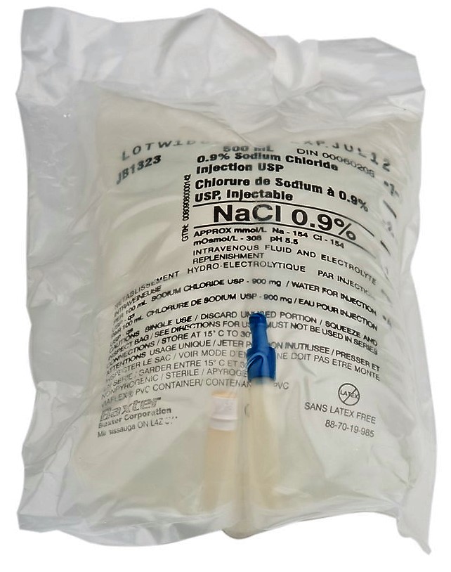
You're responding to a gunshot wound. When you arrive, you find a young man has been shot in the chest, and has significant hemorrhage. As you load him into the ambulance, your partner tells you he is spiking a 1-liter bag of 0.9% sodium chloride, also known as normal saline (NS).
You're curious, because the patient is hemorrhaging blood, not salt water, and ask why you’re not prepping blood products instead.
Your partner responds, “Because it’s something we can give him now, and it helps with circulation.”
But, does it actually help?
This review provides historical information and research with the aim of making a case against the use of NS, and why the word “normal” may be a misnomer. This can be applied to all crystalloids or clear fluid with regard to trauma resuscitation.
The goal of this article is to illustrate the many deficiencies of administering NS in a trauma patient, and to encourage critical thinking regarding current fluid resuscitation strategies that discuss increasing support for the use of blood components, including whole blood (WB).
History of Crystalloids
NS has existed in some form for nearly 200 years, largely tracing its roots back to the European cholera pandemic of 1831.1 But the solutions that were used in this outbreak, and for several decades of medicine thereafter, bear little resemblance to the modern mixture we use, both in content and in appropriate use.2
The first recorded experiment in modern IV fluid therapy is believed to be from a Russian chemist who, in treating a severely ill cholera patient, “injected 6 oz. (180 mL) of water intravenously.”1
William Brooke O’Shaughnessy, a recent medical school graduate, later published a paper in the Lancet in December 1831, stating that the goals of IV fluid in treating cholera were, “First, to restore the blood to its natural specific gravity; Second, to restore its deficient saline matters … these can only be effected … by injection of aqueous fluid into the veins.”3 Thus, we see the early foundations of saline, although referring to it as “normal” wouldn't occur until many years later.
In the 1830s, there were numerous practitioners experimenting with various solutions in an attempt to “restore the natural current in the veins and arteries” and “improve the colour of the blood.”4
In May 1832, Robert Lewins described treatments used by Thomas Latta on six patients, with solutions consisting of “two drachms of muriate, and two scruples of carbonate, of soda, to 60 ounces of water.”4 When Dr. Latta later wrote of these experiments himself, he noted a different composition of his makeshift IV fluid than was previously described.1
Other concoctions of the time included “one drachm muriate of soda, 10 grains carbonate of soda in two pounds aqua calid.”4 Historical reviews have noted that some of these compositions actually had no detailed measurements recorded at all.1
As the cholera pandemic waned in 1833, there was less urgency for IV fluid research, and for many years following, IV fluid research and publication were rare.
The first time the term “normal saline” appears in literature came decades later in the Sept. 29, 1888, edition of the Lancet.1 A patient who had “suffered over a month of vomiting, with minimal oral intake. .. [was] ... injected with 34 fluid oz. [approximately 1020 mL]” of a fluid by Churton,5 which, as shown in Table 1, bears little resemblance to the NS used today (or, with phosphate and bicarbonate, bears little resemblance to any crystalloid used today).
Table 1: Comparison of Churton's solution with normal saline
| Solution | Na+ (mmol/L) | Cl- (mmol/L) | POPO42- (mmol/L) | HCO3- (mmol/L) |
|---|
| Churton's solution1 | 150 | 128 | 2.5 | 27 |
| Normal saline | 154 | 154 | 0 | 0 |
Other isolated case reports described similar situations, but again, these IV fluid treatments weren't consistent with a 0.9% sodium chloride composition. Therefore, authors have speculated that word-of-mouth was likely to blame, rather than actual scientific research and publication.1
The NS term did find scientific support, however, after Dutch chemist Hartog Jakob Hamburger, concluded that “the blood of the majority of warm-blooded animals, including man, was isotonic with a sodium chloride solution of 0.9%,”6 effectively linking the two. As it's been described, “the scientific evidence supporting the use of 0.9% saline in clinical practice seems to be based solely on this … [otherwise] it remains a mystery how it came into general use as an IV fluid.”1
The use of NS and other crystalloids would have been used as the primary prehospital resuscitation fluid of choice for the past 30 years. However, “the historic role of crystalloid and colloid solutions in trauma resuscitation represents the triumph of hope and wishful thinking over physiology and experience.”7
Now, with this checkered background of the solution we call NS today, we present reasons why NS shouldn't be a mainstay of trauma treatment.
1. Normal saline isn't blood.
This is obvious, but it's an important introductory point. NS, as well as similar fluids like Lactated Ringers (LR), are crystalloids, and therefore consist of an electrolyte solute (in this case, sodium and chloride) suspended in a water solvent. As previously mentioned, NS solution has never truly proven itself worthy of the “normal” or “physiological” titles that it bears today.2
There are substantial disparities between the normality of human serum and NS. The comparison of NS and normal serum electrolyte ranges are listed in Table 2.
Table 2: Normal saline vs. normal serum electrolyte ranges
| Solution | Na+ (mmol/L) | Cl- (mmol/L) | K+ (mmol/L) | Ca2+ (mg/dL) | Mg2+ (mg/dL) |
|---|
| Normal serum | 134-145 | 98-107 | 3.6-5.2 | 8.9-10.1 | 1.7-2.3 |
| Normal saline | 154 | 154 | 0 | 0 | 0 |
Prior literature has described hemorrhagic shock as a type of “blood failure.”8,9 The goals of trauma resuscitation are to restore the functionality of blood, this is completed by restoring:10
- Circulating volume;
- Oxygen delivery; and
- Hemostatic potential.
NS addresses none of these critical tasks.
Prior literature has argued that NS can increase circulating volume, and that adding salt water in trauma patients will help “circulate” remaining RBCs to deliver oxygen.11 However, NS itself doesn't serve this function. It's important to acknowledge this basic, yet critical, issue, as well as its increased risk of mortality in hemorrhagic patients.12
Similarly, issues with significant extravasation that negate increasing fluid loads undermine any argument to use NS as temporary volume booster.
Simply adding fluids to a hemorrhaging body in order to theoretically “push” existing RBCs around for oxygen delivery and waste removal is largely unsupported in modern trauma literature, and, according to some, there's no human data supporting the claim that a large volume crystalloid resuscitation strategy will actually improve organ perfusion.13
Conversely, this strategy can lead to significant complications, including compartment syndrome, dilutional coagulopathy, hyperchloremic metabolic acidosis, immune dysfunction, and kidney injury.14-16
Other factors to consider include the oxygen debt and deficit that are accumulated in hemorrhagic shock. In a normal healthy body, oxygen consumption (VO2) is independent of cardiac output and therefore oxygen delivery (DO2). In hemorrhagic shock, oxygen deficit occurs when the amount of oxygen demanded by the tissues is inadequately matched by supply. Over time, these multiple deficits, result in an oxygen debt. The seriousness of an oxygen debt can't be overstated; it's the “only physiological variable that can quantitatively predict survival.”17
Crystalloids, like NS, cannot adequately repay oxygen debt in a timely manner.
2. Normal saline worsens acidosis & coagulopathy.
Most prehospital medical providers are well-versed on the three main concerns of significant hemorrhage, also known as the “lethal triad,” metabolic acidosis, coagulopathy and hypothermia. The use of NS in trauma resuscitation has been shown to exacerbate the first two aspects of this triad, metabolic acidosis and coagulopathy, as well as effect blood concentration and induce blood vessel dilation, all of which have the potential to worsen patient outcomes.18
It’s important to stress the predisposition for metabolic acidosis in trauma patients, as poorly perfused regions of tissue accumulate lactic acid and other cellular wastes, thus decreasing blood pH. Although trauma patients are already prone to acidosis, resuscitation attempts with supraphysiological chloride content in NS have been shown to worsen the patient’s condition by furthering hyperchloremic metabolic acidosis.15,18,19
The risk of NS-induced acidosis has been well-documented. One of the early reports comes from the journal Anesthesiology, where a 1997 case noted that a patient primarily received NS during the course of a long kidney surgery.20 The timeline and results progressed as follows:
- After 4 hours, the patient was estimated to have lost 1L of blood, and had received 5L NS with 1L albumin and 2 units of packed RBCs. The pH at that time was 7.28 (normal = 7.35-7.45), demonstrating an acidotic state.
- Sodium bicarbonate was then administered, and the pH rose to 7.32.
- After 8 hours, blood loss was estimated to have been 3.5L total, and the patient was noted to have received 20L of NS, with 9 units of packed RBCs and minimal other products. The pH at that point had fallen to 7.16, signaling onset of significant acidosis.
As the authors note, “the metabolic acidosis was diagnosed as a dilutional nonunion gap hyperchloremic metabolic acidosis resulting from the large volume of normal saline given during surgery and not from inadequate end organ perfusion.”20
Numerous studies have investigated the effects of NS in healthy volunteers, finding increased chloride levels and decreases in both bicarbonate and pH, thereby demonstrating the acidotic effect NS has on a healthy non-traumatized body.21,22
As this effect happens in healthy individuals, the effect in a trauma patient, already at risk for metabolic acidosis, creates an increased concern for worsened outcomes. Hemorrhaged animal models have demonstrated significant hyperchloremic acidosis from NS administration.23,24 This concern has been further validated by real-world findings of increased mortality in trauma patients when NS is used for resuscitation,13 including one large-scale study involving more than 3,000 trauma patients which demonstrated worsened outcomes when NS was used for fluid resuscitation.12
Why are there worsened outcomes with NS? What does metabolic acidosis, exacerbated by NS administration, do to the lethal triad in trauma patients?
NS-induced acidosis has been shown to directly decrease cardiac contractility and chronicity, as well as decrease the effectiveness of circulating catecholamines such as epinephrine,16,23 which can then further decrease cardiovascular function.
Furthermore, acidotic states have been demonstrated in animal models to significantly decrease fibrinogen concentration and impair thrombin generation,23,24 showing a downstream effect on other systems that are critical to stabilizing traumatic hemorrhage. Thus, many have concluded that based on NS-associated acidosis alone, the use of NS for trauma resuscitation is not supported.21
In severe trauma, effective coagulation is vital to help prevent further blood and fluid loss. Physiologically, as a crystalloid, NS likely contributes to what has been best described as trauma induced coagulopathy (TIC).9
There are two arms of trauma induced coagulopathy, acute traumatic coagulopathy (ATC) and iatrogenic coagulopathy. Overall, ATC is similar to TIC, with the significant difference being it occurs within the first 30 minutes of injury. Iatrogenic coagulopathy is the initiation or furthering of these issues through the (largely) use of improper fluid resuscitation strategies in hemorrhagic patients.
Directly, NS is linked to iatrogenic coagulopathy via the functional impairment of thrombin and fibrin, which are essential to clot formation.25 Indirectly, as mentioned above, the use of NS for resuscitation can potentiate iatrogenic coagulopathy via increased acidosis and inflammatory markers.16,23,24,26 Beyond concerns for preexisting ATC in patients with hemorrhage, a rush to administer NS likely serves to induce iatrogenic coagulopathy and thus further prevent effective coagulation.
NS contains no clotting factors or clotting support, and in fact, as it further dilutes coagulation factors and increases blood acidity, NS can significantly degrade the body’s ability to clot and achieve hemostasis.8,9Coagulopathy complications are furthered by rampant NS bolus administration in attempts to maintain or normalize BP, which could literally blast apart, or otherwise disrupt, previously clotted vessels.18
Not only does NS directly and indirectly impair new clot formation, it also has the potential to significantly disrupt and destroy existing clots. Though “lower blood pressure enhances regional vasoconstriction and facilitates clot formation and stabilization,” a patient receiving a bolus NS does the exact opposite: it uses salt water to keep vessels open and further prevents adequate clotting in areas of trauma.
3. Hemodilution & vascular changes.
As mentioned previously, a primary goal of trauma resuscitation is to achieve a stable blood pressure, and NS-led resuscitation is partially supported by the argument that it “keeps the blood circulating” for continued organ perfusion. Therefore, many protocols utilize an NS bolus of 1-2 L in an attempt to normalize SBP or MAP, as well as the quintessential 3:1 IV fluid-to-blood lost ratio that we commonly find in trauma literature.27
Although studies have proven that administration of any crystalloid fluids can have significant vasodilatory effects, this effect is greatest with NS.22,24 Research has further revealed that only 20% of infused volume remains intravascular, demonstrating a substantial loss, and waste, of administered fluid.27
NS-induced vasodilation and leakage will cause further cardiovascular stress (in addition to the metabolic acidosis-induced stress mentioned above) in order to further maintain circulatory support. Ironically, NS administration dilates and causes leakage in the very vessels it's meant to maintain, therefore producing greater stress on the systems it was given to support.
Additionally, NS has significant effects on another critical organ that's already under stress in a trauma patient: the kidneys—an important clearinghouse for metabolic wastes (including excess sodium and chloride, among others), as well as regulation of acid-base balance.
Although we referenced the vasodilation effects above, studies have established significant reductions in renal blood flow and tissue perfusion with NS administration,19 thereby harming a critical organ that helps regulate one of the very imbalances that NS creates. The "yin-yang" effect of renal vasoconstriction and vasodilation elsewhere may help explain why crystalloid administration has been determined to be an independent mortality risk factor when used in an attempt to normalize blood pressure (BP) in trauma patients that are given as little as 1.5L of NS.12
Further damage is done in the lungs, where crystalloids have been found as a modifiable risk factor in trauma and resuscitation.28
Conclusion
To summarize, the suboptimal characteristics and harmful effects of NS in trauma resuscitation include the following:
- Its inception history is vague;
- It was never initially intended for use in trauma;
- It’s not blood;
- It doesn’t mimic blood well; and
- It can actually cause significant harm and exacerbate ongoing pathology in a trauma patient, worsening their condition.
Given this, why would you administer NS in the setting of traumatic hemorrhage?
Supported by this review, it's important to reiterate and acknowledge that “the historic role of crystalloid and colloid solutions in trauma resuscitation represents the triumph of hope and wishful thinking over physiology and experience,”7 and therefore we strongly agree that “crystalloid administration should be reduced or eliminated once blood products are available.”29
Disclaimer: The views expressed in this article are those of the authors and do not reflect the official policy or position of the US Army Medical Department, the US Army Office of the Surgeon General, the Department of the Army, Department of Defense, or the US Government.
References
1. Awad S, Allison SP, Lobo DN. The history of 0.9% saline. Clin Nutr. 2008;27(2):179–188.
2. Chen L. The myth of 0.9% saline: Neither normal nor physiological. Crit Care Nurs Q. 2015;38(4):385–389.
3. O’Shaughnessy WB. Experiments on the blood in cholera. Lancet. 1831;17(35):490.
4. Lewins R. Injection of saline solutions in extraordinary quantities into the veins of malignant cholera. Lancet. 1832;18(456):243–244.
5. Churton DR. Leeds general infirmary: A case of scirrhus of the pylorus, with excessive vomiting; repeated intravenous injections of saline solution; remarks. Lancet. 1888;132(3396):620–621.
6. Hamburger HJ. A Discourse on permeability in physiology and pathology. Lancet. 1921;198(5125):1039–1045.
7. Cap AP, Pidcoke HF, DePasquale M, et al. Blood far forward: Time to get moving! J Trauma Acute Care Surg. 2015;78(6 Suppl 1):S2–6.
8. White NJ, Ward KR, Pati S, et al. Hemorrhagic blood failure: Oxygen debt, coagulopathy, and endothelial damage. J Trauma Acute Care Surg. 2017;82(6S Suppl 1):S41–S49.
9. Meledeo MA, Herzig MC, Bynum JA, et al. Acute traumatic coagulopathy: The elephant in a room of blind scientists. J Trauma Acute Care Surg. 2017;82(6S Suppl 1):S33–S40.
10. Cap AP. Whole blood (functionality): The cornerstone of remote damage control resuscitation. Oral presentation at Special Operations Medical Association Scientific Symposium; May 2017; Charlotte, NC.
11. Lilly MP, Gala GJ, Carlson DE, et al. Saline resuscitation after fixed-volume hemorrhage. Role of resuscitation volume and rate of infusion. Ann Surg. 1992;216(2):161–171.
12. Ley EJ, Clond MA, Srour MK, et al. Emergency department crystalloid resuscitation of 1.5 L or more is associated with increased mortality in elderly and nonelderly trauma patients. J Trauma. 2011;70(2):398–400.
13. Marik PE. Iatrogenic salt water drowning and the hazards of a high central venous pressure. Ann Intensive Care. 2014;4(21).
14. Van PY, Riha GM, Cho SD, et al. Blood volume analysis can distinguish true anemia from hemodilution in critically ill patients. J Trauma. 2011;70(3):646–651.
15. Santry HP, Alam HB. Fluid resuscitation: past, present, and the future. Shock. 2010;33(3):229–241.
16. Kiraly LN, Differding JA, Enomoto TM, et al. Resuscitation with normal saline (NS) vs. lactated ringers (LR) modulates hypercoagulability and leads to increased blood loss in an uncontrolled hemorrhagic shock swine model. J Trauma. 2006;61(1):57–64; discussion 64–65.
17. Barbee RW, Reynolds PS, Ward KR. Assessing shock resuscitation strategies by oxygen debt repayment. Shock. 2010;33(2):113–122.
18. Kaczynski J, Wilczynska M, Hilton J, Fligelstone L. Impact of crystalloids and colloids on coagulation cascade during trauma resuscitation-a literature review. Emergency Medicine and Health Care. 2013;1(1).
19. Chowdhury AH, Cox EF, Francis ST, et al. A randomized, controlled, double-blind crossover study on the effects of 2-L infusions of 0.9% saline and plasma-lyte(R) 148 on renal blood flow velocity and renal cortical tissue perfusion in healthy volunteers. Ann Surg. 2012;256(1):18–24.
20. Mathes DD, Morell RC, Rohr MS. Dilutional acidosis: Is it a real clinical entity? Anesthesiology.1997;86(2):501–503.
21. Schreiber MA. The use of normal saline for resuscitation in trauma. J Trauma Acute Care. 2011;70(5 Suppl):S13–14.
22. Williams EL, Hildebrand KL, Mccormick SA, et al. The effect of intravenous lactated ringer’s solution versus 0.9% sodium chloride solution on serum osmolality in human volunteers. Anesth Analg. 1999;88(5):999–1003.
23. Martini WZ, Pusateri AE, Uscilowicz JM, et al. Independent Contributions of Hypothermia and Acidosis to Coagulopathy in Swine. J Trauma. 2005;58(5):1002–1010.
24. Martini WZ, Cortez DS, Dubick MA. Comparisons of normal saline and lactated Ringer’s resuscitation on hemodynamics, metabolic responses, and coagulation in pigs after severe hemorrhagic shock. Scand J Trauma Resusc Emerg Med. 2013;21(86).
25. Sorenson B, Fries D. Emerging treatment strategies for trauma-induced coagulopathy. Br J Surg.2012;99(Suppl 1):40–50.
26. Meng ZH, Wolberg AS, Monroe DM 3rd, et al. The effect of temperature and pH on the activity of factor VIIa: implications for the efficacy of high-dose factor VIIa in hypothermic and acidotic patients. J Trauma.2003;55(5):886–891.
27. Jabaley C, Dudaryk R. Fluid Resuscitation for Trauma Patients: Crystalloids Versus Colloids. Current Anesthesiology Reports. 2014;4(3):216–224.
28. Robinson BRH, Cohen MJ, Holcomb JB, et al. Risk factors for the development of acute respiratory distress syndrome following hemorrhage. Shock. 2017. [Epub ahead of print.]
29. Dutton RP. Damage Control Anesthesia. International TraumaCare. 2005:197–201.


